My projects
Photo Video Blog Books
Goltsov Sergey
I serve the art of medical doubt

Phenotypic dermatology

The book reveals the need to move to a newly discovered form of dermatology: phenotypic dermatology. Complemented by the use of the flow cytometer and the conceptual type of evaluation of skin cell findings, dermatology has been technically and theoretically transformed. Created by synthesizing the methods of experimental dermatology, immunology, and conceptology, phenotypic dermatology allows for many practical models for the accurate diagnosis and treatment of skin diseases. The book is based on an officially confirmed discovery, the essence of which is the separation of skin cells without damaging their membrane and thus preserving their viability, which makes it possible to observe cell functions and, accordingly, to select an individual treatment for the patient. Already today we can observe the first practical results of applying the new possibilities. The book is addressed to dermatologists - from university students to professors. To the former for further development of dermatology, and to the latter for demonstration of a new, different view of the solution of old problems.


Logic of the main idea:

  1. Skin consists of cells.
  2. Cells are grouped into subpopulations.
  3. Cells of different subpopulations have different functions.
  4. The functions of cells in different subpopulations are represented by unique sets of molecules on their surface.
  5. Cells respond by activating functions in response to external and internal influences.
  6. Cell responses are summarized and expressed by changes in skin structure.
  7. Changes in skin structure are manifested by morphological elements of the rash; there are two dozen of them.
  8. Combinations of morphological elements constitute clinical diagnoses, of which 25 thousand are known in the history of dermatology.

    At this stage, the patient, being unique, is lost.
    The doctor treats the disease, but not the patient!

     
  9. Treatments for skin diseases are universal and are created without distinction of the causes that gave rise to the variety of morphological elements of the rash.
  10. By discovering the ability to separate skin cells and consider the molecules on their surface, it became clear that the variety of possible combinations of cells and their functions, and thus unique diagnoses, is much greater, specifically – 2100 663 284.
  11. By determining the functions of skin cells, using monoclonal antibodies, it becomes possible to select therapy for skin diseases individually for each patient.


On the problem of the development of dermatology

The chapter raises the relevance of the development of dermatology as a practical field of medicine. The judgment reveals the existing contradictions in dermatology that hinder its further development as a practice.


All solutions are divided into simple and... correct


With this book I will allow myself to begin a conversation about the development of dermatology and express the idea that development is characterized above all by the emergence of something new - something that did not exist before. The American philosopher Ken Wilber, in his book A Brief History of Everything, wrote: "Truth will set us free, but only if we recognize that there is more than one kind of truth. I dare say that with nihilism and denial of the new, development will not happen, this applies to dermatology as well. Especially since the need for development has long matured and is dictated by the high demand not only for individual scientific discoveries regarding a skin disease, but also for solutions to pressing problems in dermatology as a whole.

The Presidential Council for Strategic Development and Priority Projects, held in 2017, outlined the main criteria for the effectiveness of healthcare - accurate and fast diagnosis, effective treatment, patient-friendly attitude, availability, quality and efficiency of medications. Accurate diagnostics was prioritized.

At the same time, it was noted that in terms of prevalence, skin diseases in the Russian Federation in 2016 were in the top five among all diseases. The incidence of skin and subcutaneous tissue diseases was quite high both among the adult population - every 25th, and among children - every 15th child.

A total of 8,604,183 cases of skin and subcutaneous tissue diseases were reported, including 6,240,955 new cases. Skin diseases are in the top five of all diseases in terms of prevalence.

Along with this information, demonstrating the unconditional social and economic importance of skin diseases in the life of the population of any country, the development of modern dermatology does not seem possible in isolation from new technologies, through which this science, which studies the human skin, is integrated into the most complex forms of human activity. However, five years later, the prevalence and incidence rates of skin and subcutaneous tissue diseases remain high, especially in children's age groups.

At the same time, no breakthrough technologies, scientific synthesis results, or pragmatic solutions laying down novel approaches to the study of skin diseases have been found in the literature available to us in the past five years. Obviously, this is a consequence of the complexity of studying a biological system such as the human skin, which consists of a colossal number of cells and a variety of cellular relationships involved in the pathogenesis of skin diseases.

Trends in the development of science clearly demonstrate that its achievements are associated with new technologies of research̆ activities and with progressive types of thinking of the results obtained. This is dictated by the high demand for the accelerated emergence not so much of individual scientific discoveries and their engagement in practice, but rather the generation of clusters̆, classes of discoveries that form the frontline of solutions to current medical problems.

So far, dermatology, as a science, has evolved gradually, accumulating and multiplying knowledge about skin conditions and reactions in response to local and/or systemic treatments. Colossal material has been accumulated in the form of atlases, manuals, method manuals, and treatment protocols for skin conditions diagnosed on the basis of the sum of empirical observations. However, evolutionary adaptability is increasingly being replaced by revolutionary breakthroughs due to the synthesis of scientific knowledge and the state of the problem in the field.

Today, complex diagnoses of skin diseases are made by using standardized diagnostic approaches or by convening specialists in a consilium. At the same time, any doctor, with his thinking, tends to get to that place of pathogenesis, where there is the highest concentration of pathogenetic problems known to him, and the critical mass of structural and functional damages is empirically recognizable. This leaves no chance to get off the vicious path of trial and error and arrive at prognostically effective ways of diagnosing skin diseases.

The ongoing modernization of health care is focused on the development of high-tech and highly effective methods of diagnosis and treatment, as well as prompt implementation of scientific developments into practice. Dermatology, in this case, is not an exception and by 2022 the need to develop methods fully complying with the recommendations of the great Russian clinician M.Y. Mudrov, who two centuries ago called to "treat the patient, not the disease", has already matured.

Representatives of Russian and foreign schools of dermatology have already done a lot to provide objective ideas about morphofunctional features of human skin. At the same time, it should be remembered that the skin is also an independent organ of the immune system. Having a variety of cooperations of immunocompetent cells, it functions as a set of immunological reactions, which are also manifested in skin morphological changes visible to the naked eye.


Taking into account these circumstances, as well as the fact that visually discernible pictures of skin pathology are only a consequence of a complex set of intercellular interactions, studying the phenotype of its constituent cells is an urgent task of dermatology. However, due to the presence of unique skin cell connections preventing their separation and study in a viable state in vitro, this task is extremely difficult, but, as it turned out, solvable.


Human skin as cognitive complexity

The chapter contains reflections on the role of the empirical approach in the clinical thinking of the dermatologist. At the same time, the complexity of the subject, the human skin and its unique structure, is shown. As a consequence, information about the dynamics of skin disease, the degree of skin response to environmental influences, and evaluation of the effectiveness of applied external medications and cosmetics remain beyond comprehension.


Knowledge not born of experience, the mother of all validity,
is fruitless and full of errors.

Leonardo da Vinci


With documentary evidence in the form of ancient Egyptian papyri describing skin diseases and dating back to 1552 BC, as well as even earlier descriptions found in Sumerian documents 3,000 years before that, dermatology can be considered one of the oldest medical sciences.

Based on the undoubted importance of such a rich historical experience, in this book I will nevertheless allow myself a reflection that breaks with the tradition that dates back to the time of Hippocrates. There, dermatology has always been viewed as an empirical science, based on the systematization of the external manifestations of skin diseases observed by the doctor.

I will take the liberty of asserting that a tradition of observation based on practical experience has shaped this experience by a succession of subjective fallacies and refutations. Each successive generation of doctors was (and still is) given the chance to experience the diagnostic difficulties firsthand. Living out one's own experience, in order once again to correlate it with the experience of previous observations, to make their own "contribution" to strengthening the paradigm of diagnostic behavior, does not provide dermatologists with the prerequisites for further development.

Karl Popper wrote about this well in his discussion of common sense: "from repeated observations made in the past, we believe that the sun will rise tomorrow because it has done so in the past. It is taken for granted that our belief in a pattern is justified by the very repeated observations that gave rise to it."

Obviously, anyone can visually assess the condition of the skin. The doctor's assessment is more accurate than that of the average person, but it is still subjective and depends on experience, length of service, and the number of patients seen. Even in everyday cognition, people rely on previous thoughts, experiences, and generalizations to make observations. Unlike simple, everyday observations, which are mostly random and unorganized, doctor's observations are purposeful. They cannot be passive contemplation of what is being observed, since the doctor's mind not only reflects pictures of the world, but also creates them, allowing errors, delusions, and illusory perceptions to guide the doctor down the wrong path. And man is capable of convincing himself of anything.

This is well illustrated by the diagnostic algorithm used in dermatology, which is nothing more than an empirical basis denoting a body of information accumulated by dermatologists in direct interaction with objects and phenomena of the subject area under study.

There is a real multitude of disease symptoms, but one or more elements of the rash are taken into account. This does not take into account the fact that the determinacy of the origin of the rash element is different each time and comes from the unique characteristics of the patient himself.

The totality of symptoms is grouped into a diagnosis, or more precisely, diagnoses. And, metaphysically removed from the object twice, a universal treatment based on the subjectivism and empirical experience of the doctor is proposed. Everything is justified - the familiar element justifies the symptom, the symptom determines the diagnosis, and the latter, through clinical recommendations, justifies the prescription of drugs.

Such assumptions are of an orderly nature, assembled into diagnoses. For this, however, the dermatologist needs experience and time to form a specific image for each diagnosis, which means that dermatology in this sense is an empirical science, and we know that the basic attribute of science is the ability to predict, not the transmission of experience. The Scottish philosopher David Hume wrote well about this, that "repetition has absolutely no evidentiary power, though it plays a dominant role in life and 'understanding. He wrote about this nearly three centuries ago. It is strange that we have not yet learned the lessons.

It is well known that the dermatologist knows two dozen signs - these are the primary and secondary morphological elements of the rash. Their successive transformation and combination into symptoms forms the diagnostic field of the dermatologist, expressed by twenty-five thousand diagnoses. Of course, this diversity forces the dermatologist to group the diagnoses under the principles of treatment he knows.

Thus, when tested asking for a diagnosis, 7 out of 10 dermatologists I knew with more than 10 years of clinical experience could not even suspect the diagnosis of dermatomyositis. What could be easier, though? Erythema oedemas of a wine-red color in the face, neck, back of the hands, and knees may be the only manifestation of this disease for a long time. In this case, a typical periorbital "spectacled" erythema is presented, due to which the face has acquired a "weeping" expression in combination with typical symmetrical skin lesions of the trunk and extremities.

Two of the remaining three suggested a generalized form of rubromycosis, and one doctor even suggested toxoderma. And, of course, it is possible to assume visually these diagnoses as well, requiring additional confirming or refuting studies.

A patient diagnosed with dermatomyositis.

It turns out that the morphological elements of the rash, designed to streamline diagnostic thinking, become the keys to the door of diagnostic uncertainty. By their ease of use as primary diagnostic criteria, they add to the doctor's already existing need for patterns that explain further actions (including treatment prescription). Often, this dependence forces one to find patterns even where they do not and never have existed.

Оттого, что дерматология продолжает, спустя тысячи лет, опираться на визуальную диагностику возникает следствие – раннее использование средств симптоматического подхода к лечению. Дерматология движется в русле поиска однотипных лечебных средств для решения разных проблем.

Использование медицинской лупы и эпилюминисцентной дерматоскопии продиктовано потребностью доктора увидеть больше и познать элемент сыпи тщательнее в поиске дополнительных критериев различения. В подтверждение своих слов, демонстрирую два случая клинического наблюдения.

You can tell from the shoulder-blade articulation that these are two different people, but do they have the same diagnosis? It is very similar, isn't it? Visually and anamnesthetically, both patients demonstrate a similar visually observed clinical picture. Dare we say they are indistinguishable. But, is this true?

Guided by the classical scheme of making a clinical diagnosis based on the rash elements visible to the eye, we can talk about many diagnoses (lymphoma, erythroderma, psoriasis, eczema, toxicoderma...) and not only these. But, the treatment approaches and prognosis are different, aren't they?

This is what happened. The essentially identical treatment approach resulted in Patient 1 demonstrating a rapid improvement in his skin condition and was discharged from the hospital in 2 weeks, unlike Patient 2, who died 4 months after admission.

The subjective-visual level of diagnosis has long been in need of information at the level of cells and molecules, which opens up the space of clinical conditions that have not yet been named. They are as numerous as the skin cells with their functions performed within the organ.

Being a two-component tissue system formed by epidermis and dermis, the skin is represented by a number of subpopulations of cells with a strict functional purpose of their functions, which is reflected in the skin's ability to act as a barrier between the human external and internal environments.

Forming a vast area of contact with the external environment and being the most important barrier tissue limiting the internal environment of the organism, the human skin has historically formed as an independent organ of the immune system, often being the main bridgehead for implementation of most of its response mechanisms.

Besides, having a variety of immunocompetent cells cooperating with each other both with the help of complementary structures on the surface and with the participation of immunoregulatory cytokines, testify to the fact that skin is not only an active immune organ, in which resident and recurrent epidermis and dermis cells can initiate inflammatory processes both in the whole body and in situ.

The cellular substrate of skin immune competence is represented by resident (mast cells, Langerhans cells, keratinocytes, endothelial cells, fibroblasts, monocytes (macrophages) and recirculating (lymphocytes and granulocytes) cells of medullary origin.

Thus, skin immune system combines the features of local functioning and integration into the general immune system of an organism. Dynamics of the skin functional state, ensured by constant renewal of the composition of recirculating lymphocytes between regional lymph nodes, blood and skin, thus creating immune homeostasis, causes difficulty in studying properties of cell subpopulations, but simultaneously opens up an opportunity to comprehend immune mechanisms, so far considered by dermatologists only theoretically, mainly in conditions of chronic inflammation.

The fact that skin is an active immune organ is evidenced by a highly efficient set of cellular and humoral elements, in which resident and recurrent cells of epidermis and dermis are capable not only to initiate immune processes, but also to participate in them.

This leads to a variety of immunopathological reactions and is expressed in a huge variety of variants of the clinical picture of various skin diseases, which creates a clear cognitive difficulty for the dermatologist during examination of the skin.

Lack of pathogenetic information about morphological manifestations of skin diseases makes dermatologists simplify the observed phenomena to the known diagnoses, justifying diagnosis and treatment of skin diseases only by visualization of the latter. Often, when making a diagnosis, the dermatologist uses abstract concepts known to him, the relations between which only approximately reflect the relations between real skin cells and membrane events on their surface.

Undoubtedly, the diagnoses are important already because they relatively correctly, but still reflect the properties and relations of the elements of the examined populations. This makes it possible to describe the observed picture and, with the help of language, to carry out the accumulation and transfer of knowledge.

When discussing the visible part of inflammatory processes and the pathogenetic mechanisms underlying their occurrence, we should consider them as parts of a single whole. More precisely, as a system of heterogeneous objects united into one whole - the human skin. Functionally interacting with each other, these parts determine the entire variety of rash elements and their transformations observed by dermatologists.

Visible rash elements - morphological elements, as well as the skin as a whole, qualitatively change their state as a result of changes occurring at the level of cells and surface active molecules expressing the function of these cells. The latter, for the objectivity of judgments about the diagnosis, should be described in quantitative terms.

At the same time, each skin cell can be considered as a separate integrity consisting of its own parts and membrane events on the surface determining its phenotype, each of which can be investigated separately. This is for us to consider in more detail.

Relevance of flow cytometry application for evaluation of phenotypic diversity of skin cells

The chapter argues that numerous known methods of skin research do not solve the issues of differentiation of skin conditions at the cellular level, and this does not allow dermatologists of the present day to fully assess the functional activity of skin cell subpopulations under normal and pathological conditions. This forms the challenges to dermatology, which allowed the author to formulate a number of questions, impossible to raise earlier. On this basis, the scientific problem, object, subject and purpose of the study are shown.

Dermatology is a rational philosophy about the skin.


For a long time, the only way to obtain objective information about the morphofunctional state of skin cells remained incisional biopsy, the invasiveness of which limited its application and practically excluded dynamic observations.

In their turn, noninvasive methods of skin examination - corneometry, seboumetry, cutometry, profilometry, and for deep investigations - optical coherence tomography, ultrasound microscopy, magnetic resonance imaging, describing a number of skin properties, do not provide an opportunity to assess a complex complex set of intercellular interactions. For all their persuasiveness in solving individual tasks of dermatology, they do not provide reliable information about functional differences of skin cells in quantitative terms. Describing a number of skin properties, the listed methods do not take into account the fact that the skin is an organ of the immune system, which is constantly influenced by the environment and in which a complex set of intercellular interactions is constantly taking place.

In fact, the extensive development of dermatology has formed adherents of two extremes in skin research. Some authors, relying on the possibility to study the skin and its functions with noninvasive methods, deprive themselves of information at the cellular level, where a huge amount of information about the nature of inflammatory reactions opens up. Others actualize invasive methods, which, although they allow to study the skin at the cellular level, do not provide an opportunity to evaluate the function of cells, because they work with fixed (dead) tissue, which is known to be functionally inactive.

The observed state of dermatology, aggravated by the increasing variety of skin diseases, comorbidity and lagging rate of development of diagnostic methods, is a reflection of the weakness of dermatology as a theory. Within the current paradigm of dermatology, which relies on imaging diagnostics, reticent and limited in its use of advanced cellular diagnostic methods, no new conceptual problems are emerging that would act as motivators for its development, not just improvement. Dermatology is moving in the direction of the search for single-type therapeutic agents universally satisfying the practice. Obviously, this brings improvement in performance, but it also indicates that the paradigm itself is exhausted. 

Thus, modern dermatology, first of all as a science, has an urgent need for intravital methods of morphofunctional skin examination, and visual examination is still the main method of skin condition assessment.

Indistinctness of skin conditions at the cellular level and assessment of cells fixed by histological methods does not allow dermatologists of the present day to fully assess dynamics of skin disease, degree of skin response to environmental influences, functional activity of skin cell subpopulations in normal and pathological conditions, to objectify criteria of skin age changes and evaluate effectiveness of applied drugs and cosmetics.

This circumstance makes it relevant to study the phenotype of skin cells, the knowledge of which will strengthen the diagnostic tools of the dermatologist approximately as it happened at the time of microscope discovery. All the more, the dynamics of increasing variety of skin diseases is much higher than the rate of active use of new diagnostic methods.

At the same time, technologization and development of such fields of medicine as immunology and cytology stimulate technological renewal of dermatology.

Thus, based on the method of flow cytometry it became possible to determine not only quantitative parameters of cell populations, but also their broader subpopulation composition, assessing with high precision the functional properties of the cells of the immune system. It turned out that not only hers. Discovery of a method to obtain skin cytoimmunogram, reflecting state and functionality of its cells, gave rise to the possibility of detailed study of cell subpopulations not only in peripheral blood, but also in skin.

At the current stage of scientific development, direct observation of membrane events on the surface of skin cells is not feasible, but they can be judged by flow cytometer readings, which can be admissibly considered as observable values. The use of a high-precision device makes it possible to exclude errors and subjectivity of observations. This will make it possible to introduce into dermatology an accurate substantiation of diagnoses based on the observed phenomena and thus make a dialectical leap from the empirical form of skin disease diagnostics to the rational-theoretical one. 

Considering that the skin functions through the specialization of cell subpopulations, and that visually distinguishable patterns of skin diseases are only a consequence of a complex set of intercellular interactions, studying the phenotype of the cells that form it is an urgent task of dermatology. It is also obvious that true development of dermatology is not possible in isolation from cell technologies. At the same time, cell technologies exclusively stimulate renewal of dermatology, which allows to thoroughly study the processes of formation of a variety of skin cell phenotypes, reflecting their specialization and selectivity of functions.

Thus, phenotyping by flow cytometry allows to characterize cells using monoclonal antibodies and to judge their functional state by the presence of cell markers. Two essential features make this method particularly valuable for practice:

Characterizes heterogeneous cell populations by phenotype;
Detects and characterizes events occurring at a frequency of 10-5 to 10-7.
Flow cytometry readings allow to judge cell morphological characteristics (size, nucleus-cytoplasm size ratio, cytoplasm granularity, degree of cell asymmetry), determine subpopulation composition and assess their functional state with high accuracy. Discovery of a method to obtain skin cytoimmunogram, reflecting the condition and functionality of its cells, gave rise to the possibility of detailed study of cell subpopulations not only in peripheral blood, but also in skin.

Scheme of the flow cytometer operation.

Experimental construction of skin cytoimmunograms is performed using an invasive method involving skin biopsy sampling to a depth of 2 mm using a 2 mm panch biopsy tool. The obtained skin sample can be examined ex tempore or after cryopreservation. The cell suspension is phenotyped by flow cytometry on a Beckman Current Cytomics FC500 flow cytometer. The technical details of the method will be thoroughly discussed in the next chapter.

Beckman Current Cytomics FC500 Flow Cytometer

As an integral tool for determining the phenotype of tissue cells based on the principle of antigen-antibody binding detection, specific antibodies can be obtained for almost any protein (or its fragment), which means that immunophenotyping can be used in almost any field of medicine, dermatology being no exception. All the more so because flow cytometry is a reliable method for rapid analysis of cellular activity. This technology provides a clear picture of cell behavior and a better understanding of cellular response mechanisms to specific influences, both in normal and pathological conditions.

Since resident cells are known to play a major role in skin homeostasis, the choice of marker type is determined by the research task, which requires identification of skin cell subpopulations most influential in the pathogenesis of the disease under study.

Markers to monoclonal antibodies produced by skin cells belonging to different cell clones, i.e. originating from different progenitor cells. Monoclonal antibodies can be produced against any natural antigen that the antibody will specifically bind. As such, they can be used to detect (detect) them on the surface of the membranes of the cells that express them. 

As early as the beginning of the twentieth century, Paul Ehrlich postulated that if a component capable of selectively binding a disease-causing component could be produced, then a toxin could be delivered to the pathogen along with this component.

In the 1970s, though, tumor B-lymphocytes (myeloma cells) were already known to synthesize the same type of antibodies. These cell cultures were used to study the structure of the antibody molecule, but there was no technique to produce an identical antibody to a given antigen.

The process of producing monoclonal antibodies was invented by Georges Köhler and César Milstein in 1975. For this invention they received the Nobel Prize in Physiology in 1984. The idea was to take a line of myeloma cells that had lost the ability to synthesize their own antibodies and fuse such a cell with a normal B-lymphocyte synthesizing antibodies, in order to select the resulting hybrid cells synthesizing the desired antibody after the fusion. This idea was successfully implemented and by the early 1980's commercial production of various hybrids and purification of antibodies against given antigens had already begun.

For antigen recognition in 1982 the classification of clusters of differentiation (CD for short), a nomenclature of human differentiation antigens for the identification and study of surface membrane proteins, was proposed. CD-antigens (or CD-markers) can be proteins that serve as receptors involved in cell-to-cell interactions and are components of certain signaling pathway cascades. However, they can also be proteins performing other functions (e.g., cell adhesion proteins or apoptosis proteins). The list of CD antigens in the nomenclature is constantly growing and currently contains over 350 CD antigens and their subtypes. 

Why this magnificent discovery has not yet found practical application in dermatology remains a mystery. This is particularly paradoxical in light of the fact that the flow cytometer allows one-by-one counting of cells and their expressed membrane molecules in a population of millions of cells for their size or function - activation, proliferation and dynamic phenotypic changes - with an information collection rate of up to 1000 events per second in real time, taking only 1 sample from a patient.

Based on the scientific information that keratinocytes make up more than 90% of the cells of the upper skin layer, the epidermis, and dermal cells are overwhelmingly represented by fibroblasts (fibrocytes), mast cells, monocytes (macrophages), endothelial cells, dendritic cells and lymphocytes, of which 90% are T-lymphocytes located in the upper layers of the dermis and epidermis, and 10% are B-lymphocytes located in the middle and deep layers of the dermis, the following were taken as a basic set of markers differentiating cell subpopulations and characterizing the dynamics of membrane events in these subpopulations: CD3, CD4, CD8, CD14, CD16, CD19, CD34, CD44, CD45, CD49, CD54, CD63, CD80, CD146, CD203c; CD207, CD249.

The technical capabilities of flow cytometry to analyze subpopulations of cells and identify any number of them, as well as measure their surface and intracellular markers, assessing their functional status, together with the demand by dermatologists for a more rigorous understanding of pathological processes at the cellular level, oblige the methodological standardization of diagnostic capabilities and approaches to measuring the variety of functional states of skin cell phenotypes for diagnostic and treatment

This has already led to new questions for dermatology, to which the existing scientific paradigm does not provide answers:

  • What is the condition of the skin indicated by the particular cell compositions of all species in a particular sample of its biopsy?
  • What is the complete diversity of cell signatures of all species?
  • What combinations of features of cells of each species create particular functions in the skin?
  • What is the full composition of possible skin cell functions that arise in a variety of combinations of all cell signatures of all species?
  • What functions do particular cell species have when activated by their particular lobes in a particular skin biopsy?
  • Can a cell of each species act simultaneously in functionally different states and in which states?
  • Are there any restrictions on the occurrence of cell subpopulations that form its different cellular phenotypes?
  • What are the properties of cellular phenotypes formed by combining certain fractions of cells of each species with their functions?
  • What structure of cells, their traits, functions, and distribution of cell fractions should be taken as a "unit" of skin condition for its pragmatic use in the research and treatment practices of future dermatology?

As the space of these questions expands, there are objective difficulties in using knowledge of the diversity of functional states of skin cell phenotypes in diagnostic and treatment practice, approaches to measuring this diversity, and approaches to developing treatments. 

These and similar questions expose a problem that is formulated as follows: how is it possible to see and understand the picture of skin states that opens up at the level of the study of its cellular phenotypes, with the practical and theoretical use of knowledge about the consequences of the dynamics of its cellular diversity?

This problem is a problem of conceptual significance, in relation to which it is not yet clear how to think about it in order to solve it. In this area of research practice, what might be called a deficit of concepts for distinguishing and explaining the new reality turns out to be essential. This circumstance prompts the formation of a front of new concepts concerning skin conditions and the search for methods of generating meanings for a new type of dermatology - phenotypic dermatology.

The solution of the indicated scientific problem, turning to the cellular level of research of skin conditions, is conceived on the basis of the tools of a scientific-technical direction, founded by the Russian scientist in the field of organizational management and methodology of information system development Nikanorov S.P. and conceptual analysis technologies built on its basis. All of them are focused on the construction and synthesis of concepts in those areas of practice, which need conceptually rigorous distinctions of reality. All of them are oriented to construction and synthesis of concepts in those fields of practice, which need conceptually strict distinctions of reality.

The application of conceptual analysis that is constructively and wholly focused on obtaining practically and theoretically meaningful results will allow the theoretical development of the results of many years of human skin research. It is especially relevant in the processes of modernization of dermatology all over the world.

That is why the object of research is defined by human skin conditions at the level of its cell subpopulations; the subject of research is the properties of human skin cell subpopulations determining phenotypic diversity.

Karl Popper argued that "we never start with observations, but always with problems - either practical problems or problems of a theory that has run into difficulties. By critically discussing and testing these hypotheses, we exclude those that solve our problems worse, and choose better ones that solve them more effectively and adequately. "In doing so," Popper writes, "I draw on the neo-Darwinist theory of evolution, but in a new formulation in which 'mutations' are interpreted as a method of more or less random errors, and 'natural selection' as one of managing them by eliminating errors."

The goal in the present work is defined specifically and serves to eliminate the discrepancy between the new facts and the old ways of explaining them, namely, the conceptual justification of applying flow cytometry to assess the phenotypic diversity of human skin cells.

In its totality, all of the above became the basis for further theoretical assumptions and practical implementations. 

Method for assessing the subpopulation composition of skin cells and method for obtaining skin cytoimmunograms

The chapter briefly describes the history of the discovery of the skin cytoimmunogram and details the method of obtaining it. The practical application of the discovery demonstrates the need for profound changes in diagnostic and therapeutic approaches in dermatology as a scientific discipline and practice. 


The arrow that hits its target flies forever.
Nabokov V.V.


The ancient Greek word method denotes a way to achieve a goal, which in this work was defined as an attempt to conceptually justify the application of flow cytometry to assess the phenotypic diversity of human skin cells. Achievement of this goal will help to solve the scientific problem of building a theory explaining skin conditions at the level of phenotypes of its cell subpopulations, suitable for practical and theoretical development of dermatology and pragmatic use of knowledge about the consequences of dynamics of phenotypic diversity of human skin cells.

G.I. Ruzavin, in his book "Methodology of Scientific Cognition", by method means an ordered and organized way of activity aimed at achieving a certain practical or theoretical goal. The invention of the method is initially associated with the solution of specific practical problems through the performance of a number of labor operations, guided by the appropriate techniques, means or methods in a strictly defined order.

"By method," Descartes writes, "I mean precise and simple rules, the strict observance of which always prevents the false from being mistaken for the true - and, without unnecessary waste of mental energy - but by gradually and continuously increasing knowledge, contributes to the mind reaching a true knowledge of all that is available to it. He recommends three rules of method as basic requirements: 1) begin with the simple and obvious; 2) from it by deduction derive more complex statements; 3) act in such a way that not a single link is missed, i.e., keeping the chain of inferences unbroken.

Descartes' ideas about the deductive nature of the method of science were further developed on a broader basis by H.W. Leibniz, who sought to reduce reasoning to computation, so he became the forerunner of modern symbolic, or mathematical, logic.

With these theses I want to lay down some rigor in further judgments. Especially necessary when interpreting the results obtained.

"The regularities directly verifiable by experiment do not change. Of course, changing them is conceivable or logically possible, but this possibility is not taken into account by empirical science and does not affect its methods. On the contrary, the scientific method assumes the invariability of natural processes, or 'the principle of the uniformity of nature.'"

The history of dermatology and cell biology "remembers" a mass of attempts to separate the cellular substrate of the skin to obtain a suspension and study the phenotype of the cells comprising it. However, the method of obtaining a viable heterogeneous skin cell population was discovered and patented only in 2013. 

Report "Prospects of application of skin cytoimmunogram". Associate Professor Goltsov S.V.

Research results showed the possibility of obtaining cell suspension with viability in native samples up to 99.8%, and after cryopreservation samples up to 87.0%. The discovery was recognized as promising for the development of dermatology and practical for treatment of skin diseases. The resolution of the "X International Conference of Immunologists of the Urals" recommended the patent for implementation in the public health system.

Presidium of X Conference of Immunologists of the Urals with International Participation, dedicated to the memory of S.N. Teplova, Honored Scientist of the Russian Federation, Professor, M.D., Tyumen, 2012 E.A. Kashuba, Rector of Tyumen State Medical Academy, Professor, M.D.; N.S. Brynza, First Deputy Director of Health Department of Tyumen Region, Head of Department of Health Organization and Public Health of Tyumen State Medical Academy, Professor, M.D.; V.A. Chereshev, Academician of RAS and RAMS, President of Russian Scientific Society of Immunologists and Ural Society of Immunologists, Allergologists and Immunorehabilitationists, Director of Institute of Immunology and Physiology UB RAS, Chairman of State Duma Committee on Science and High Technology, Professor, Doctor of Science; V.P. Melnikov, Academician of RAS, Director of Tyumen Scientific Center SB RAS, Professor, Doctor of Geography; I.A. Tuzankina, Scientific Secretary of the Institute of Immunology and Physiology, UB RAS, Professor, Doctor of Science; A.A. Yarilin, Professor, Doctor of Science.

Scientific activity, through the methodology of cognition, considers the real picture of the world and serves as the main way of transformation of scientific knowledge into practical activity. It is realized through the invention of methods of scientific research, or (as it took place in our study), improvement of the existing one.

Immunophenotyping by flow cytometry allows the characterization of cells using monoclonal antibodies and makes it possible to judge their type and functional state by the presence of a particular set of cell markers and the processes occurring in them. As a method of measuring cell characteristics, flow cytometry emerged from the synthesis of knowledge of traditional histochemical and cytochemical methods of analysis. Created to accelerate analysis in clinical cytology and cytodiagnostics, this technology has gradually evolved into an effective approach for many important tasks in cell biology, immunology, cell engineering, etc.

The information extracted by flow cytometry allows to judge the morphological characteristics of cells (size, ratio of nucleus and cytoplasm size, granularity of cytoplasm, degree of cell asymmetry). But further development of technology led to the fact that researchers have in their hands such a tool as monoclonal antibodies, which provided an opportunity to type cells not only due to their morphological differences, but also due to the set of surface antigens and receptors, characteristic for certain cells and their functional state. 

Simultaneously with the familiarization with the possibility of the method, challenges to dermatology were identified, coming from the complication of skin pathologies, accompanied by significant changes in skin cells and their functions. As it turned out later, this is expressed both in changes in the number of cells and in the appearance on their surface of certain functional molecules, in their totality characterizing certain clinical conditions.

Preparation of a skin cell suspension


Experimental construction of skin cytoimmunograms is carried out by invasive method involving skin biopsy sampling to a depth of 2 mm from human buttock area using DERMO-PUNCH 2 mm biopsy instrument (STERYLAB, Italy).

Punch biopsy is a method of skin sampling using a special tubular scalpel - punch (from English punch - hole puncher, composter). The method allows you to obtain a cylindrical tissue sample of a given height and diameter. When the material is taken, the punch is directed perpendicularly to the surface of the skin, stretching the skin along the sides. At the same time, the punch is lightly pressed from top to bottom and rotated clockwise and counterclockwise until it sinks to the desired depth. Afterwards the punch is gently extracted. The biopsy specimen is extracted from the scalpel cavity with a needle and the lower part of the specimen is separated from the patient's body using sharp scissors or a blade. If necessary, sutures and a bactericidal plaster are applied to the resulting wound.

The skin biopsy and 1 ml of 0.9% aqueous sodium chloride solution are placed in the working chamber of an automatic system for mechanical homogenization of tissue Medimachine (Becton Dickinson, USA).

After that, the homogenate is filtered through an inert cell filter Falcon (Becton Dickinson, USA) with a nylon mesh structure and a pore diameter of 20 µm.

The technical result is achieved by invasive sampling of a human skin biopsy sample using a DERMO-PUNCH 2 mm biopsy instrument (STERYLAB, Italy).

Then a skin biopsy sample and 1 ml of 0.9% aqueous sodium chloride solution is placed in the working chamber of the Medimachine automatic system for mechanical tissue homogenization (Becton Dickinson, USA).

After that, the homogenate is filtered through an inert cell filter Falcon (Becton Dickinson, USA) with a nylon mesh structure and a pore diameter of 20 µm.

The tissue is homogenized for 30 seconds at +23°C. The homogenate is extracted with a sterile syringe. The working chamber of the homogenizer is washed three times with 1 ml of 0.9% aqueous sodium chloride solution. The homogenate is filtered through a Falcon cell filter (Becton Dickinson, USA) with a nylon mesh structure and a pore diameter of 20 µm. The homogenate was then centrifuged to remove supernatant liquid at 400 g for 5 min at +23°C.

After that, cell viability was determined using the intracellular dye 7-amino-actinomycin D RUO (7AAD) (Beckman Coltrer, USA). The analysis is performed on a Cytomics FC500 flow cytofluorimeter (Beckman Сolter, USA). Cell identification is performed by recording two parameters: side scatter and fluorescence registration by channel 3 (FL3).

The obtained skin sample can be examined ex tempora, or after prolonged cryopreservation. For the second option, the sterile-checked sample is placed in a 2 ml Costar cryo-tube with freezing solution (90% Fetal Bovine Serum and 10% DMSO as cryoprotectant), then the sample is frozen in liquid nitrogen vapor t°-140°C at 1°C per minute, by vitrification.

Programmed freezer.

In either case, for the first, native, or the second variant (after thawing), skin cells were incubated for 20 minutes in a light-protected place with monoclonal antibodies conjugated with fluorescein isothiocyanate (FITC), phycoerythrin (PE), PE - Texasred (ESD), PE/CY5(PC5), PE/CY7(PC7) (Beckman Coltrer, USA) fluorochromes.


Skin cell phenotyping


Using flow cytometry the cell suspension was phenotyped on a Cytomics FC500 (Beckman Colter, USA) using specific markers: CD3, CD4, CD8, CD14, CD16, CD19, CD34, CD44, CD45, CD49, CD54, CD63, CD80, CD146, CD203c; CD207, CD249, which identified cell subpopulations and determined their phenotype.

Since resident cells are known to play a major role in cutaneous immune homeostasis rather than those attracted from the circulation, the choice of the type and amount of fluorescent dyes was determined by the task of the study for which it is necessary to identify subpopulations of skin cells that most affect the pathogenesis of commonly encountered skin diseases.

Phenotyping of skin cells was performed using specific markers

Knowing that keratinocytes account for over 90% of cells in the upper skin layer, the epidermis, and dermal cells are overwhelmingly represented by fibroblasts (fibrocytes), mast cells, monocytes (macrophages), endothelial cells, dendritic cells and lymphocytes, of which 90% were T-lymphocytes located in the upper layers of the dermis and epidermis and 10% were B-lymphocytes present in the middle and deep layers of the dermis, sets of cell differentiation markers were determined to best characterize the state dynamics of these subpopulations.

This solution allowed to improve the flow cytometry method up to the possibility to obtain estimates of quantitative and qualitative composition of individual subpopulations of skin cells:

CD49f+ keratinocytes, of which CD49f+HLA-DR+ activated. Located in the epidermis, cells of ectodermal origin whose intermediate filaments are represented by keratin protein, keratinocytes make up to 90% of the skin epidermis in all mammals, including humans. Differentiated and keratinocytes of the skin epidermis are highly heterogeneous.
fibroblasts (fibrocytes) CD45-CD14-CD44+, of which activated CD45-CD14-CD44+CD80+. As connective tissue cells synthesizing extracellular matrix and located in the dermis, fibroblasts secrete precursors of collagen and elastin proteins as well as mucopolysaccharides. Fibroblasts and fibrocytes are two cell states, the first in an activated state, the second in a less active state associated with vital processes and tissue metabolism. Currently, there is a tendency to call both forms fibroblasts. Fibroblasts, just like epidermal keratinocytes, are morphologically heterogeneous with different manifestations of function depending on their location and activity. CD44 is a glycoprotein that plays an important role in intercellular interactions, cell adhesion and migration and is involved in various cellular functions such as lymphocyte activation, recycling and homing, hematopoiesis and tumor metastasis.
CD249+ mast cells, of which activated CD249+CD63+ cells, are highly specialized immune cells of connective tissue, analogues of blood basophils, also involved in adaptive immunity, specifically mediation of cytotoxic effects. Mast cells play an important role in inflammatory reactions, particularly allergic reactions. Like basophils, the surface of mast cells has receptors for IgE. When activated (e.g. in an allergic reaction), mast cells release their granule contents (heparin and histamine) into the surrounding tissue (degranulation). The release of these substances leads to changes in the intercellular substance of the connective tissue, the hematotissue barrier.
CD45+CD14+ monocytes (macrophages), of which CD45+CD14+HLA-DR+ activated. Located in the dermis, monocytes originate from hematopoietic stem cell progenitors in the bone marrow. Circulating in the bloodstream for one to three days, they then usually move into tissues throughout the body, including the skin. The mature monocytes then differentiate into resident macrophages or dendritic cells. Both monocytes and macrophages are phagocytes, acting as non-specific defense factors (innate immunity) and also to help initiate specific defense mechanisms (adaptive immunity). Monocytes and macrophages phagocytize (absorb and then digest) cellular debris and pathogens, or as stationary or mobile cells stimulate lymphocytes and other cells of the immune system in response to the pathogen.
CD207+ intraepidermal macrophages (Langerhans cells), of which CD207+CD80+, CD207+HLA-DR+, CD207+CD80+HLA-DR+ are activated. This is a subpopulation of dendritic cells of bone marrow origin located in the epidermis, accounting for 24% (other reports 38%) of all epidermal cells. Located in the epidermis, Langerhans cells represent a unique subset of dendritic cells located in the epidermis. Found in epithelial tissues, CD207 is a transmembrane glycoprotein used as a unique marker of Langerhans cells. Although these cells were described as early as 1868, an in-depth study of their morphological and functional properties is associated with the establishment of their important immunological functions in the skin. Intended to receive, process and present information about environmental antigens as well as endogenous antigens, they belong to the macrophage-monocyte-histiocyte lineage and play a central role in immune response initiation.
CD146+ endothelial cells, of which CD146+CD34+, CD146+HLA-DR+, CD146+CD54+, CD146+CD54+HLA-DR+ activated. Endothelial cells located in the dermis and lining blood vessels have an amazing ability to change their number and location in accordance with local features of skin functions. Participating in blood circulation organization, these cells create a flexible adaptive life-support system with ramifications in all areas of the body, including the skin. If it were not for this ability of endothelial cells to expand and repair the network of blood vessels, tissue growth and repair processes would be impossible. The CD146 marker is used to identify embryonic mesenchymal stem cells. Its expression can be associated with multipotency of mesenchymal stem cells, and detection indicates a high differential potential.
CD34+CD45dim epithelial progenitor cells;
lymphocyte populations: CD45+CD3+ T-lymphocytes, CD45+CD3+CD4+CD8- T-helpers, CD45+CD3+CD4-CD8+ T-suppressors, CD45+CD3-CD19+ B-lymphocytes, CD45+CD3-CD16+CD56+ NK-lymphocytes. 

Registration and interpretation of results


To record the results obtained, a "Skin Cytoimmunogram" medical document form was developed, which presents the composition of skin cells, indicating the phenotype of each subpopulation for a laboratory technician to fill in numerical data in relative and/or absolute units according to the results of the study.

The upper part of the document contains the logo of the medical institution, service sign, mailing address, contact telephone numbers, website of the medical institution, as well as free areas for the laboratory assistant to fill in the skin cytoimmunogram identification number with the date of analysis, the patient's last name and age.

In the middle part of the document, the composition of skin cells is presented in two columns, indicating the phenotype of each population. In front of each group of skin cells there are empty fields in the form of double rectangles for the laboratory technician to fill in the numerical data in relative and/or absolute units according to the results of the study. Presented numerical data indicate the number of skin cells of a particular phenotype.

In the lower part of the document, there are free areas intended for the doctor to fill in information about the results of skin cell phenotype examination, which may indicate: dynamic assessment of the course of the disease, effectiveness of the use of prescribed drugs or cosmetics, assessment of age-related skin changes, individual selection of medications, assessment of the degree of skin cell response to certain effects. Also filled out by the lab technician in the free sections of the information about the performer and the doctor who sent to conduct the analysis.

In the perspective of large-scale application, this method of skin research will make it possible to create a sex-and-age register of skin condition, objectively assess the dynamics of skin diseases, individually select medications, monitor the effectiveness of external medications, form criteria of age-related skin changes, objectively indicate the current status of patient's local immunity and may become a substantial basis for treatment and preventive programs in dermatology.

Results of immunological research of skin cell subpopulations reveal a picture which modern dermatology has no clear relation to yet. But, the translation into effective remedies of the first useful phenomena of this picture already indicates that a new history is beginning in this scientific discipline.

Practical significance of skin cytoimmunogram

The chapter briefly describes the history of the discovery of the skin cytoimmunogram and details the method of obtaining it. The practical application of the discovery demonstrates the need for profound changes in diagnostic and therapeutic approaches in dermatology as a scientific discipline and practice. 

True philosophy has to be practical

Teslinov A.G.

Translated from Ancient Greek, pragmatics, meaning "deed, action", studies the relationship between sign systems and those who use them. Highly estimating the procedure of measurements the outstanding Russian scientist D. I. Mendeleev (1834-1907) wrote: "Science began when people learned to measure; exact science is unthinkable without measures". But measurements are never perfectly accurate, no matter how carefully an experiment or experience is conducted. The inaccuracy of the results of an experiment or experience and, therefore, the relativity of natural scientific truth is due to two factors: objective and subjective. Especially since the standard method of verification of any discoveries is direct experimental verification, as they say, experiment is the criterion of truth.

Nature, in realization of its mechanisms, on the way of evolution, gives us an opportunity to formulate general, universal laws on the basis of study of its separate parts, by using synergetics of knowledge, as it took place in our case, through synergism of dermatology, cytology, immunology to enter the pragmatic study of human skin and its constituent cells. It is at the junction of different specialties that we have obtained new knowledge satisfying the practice in answering the questions and practical difficulties that have arisen.

Thus, at the solution of the set tasks and verification of a scientific hypothesis in the work a complex of methods of research complementing each other was used: method of 3D-visualization and measurement of skin structures, methods of sebumetry, corneometry, cutometry, profilometry, punch-biopsy, mechanical tissue homogenization, method of flow cytometry, method of tissue cryopreservation, method of system analysis, method of conceptual clearing of complex subject areas, method of double-knee ascent from empirical concrete to theoretically concrete through abstract, method of generic explication of concepts, methods of statement calculus and others.

Their use in research activities allowed to develop conceptual foundations of the theory of phenotypic diversity of human skin cells, providing an intensive transition to the development of research and treatment practice in dermatology. This was made possible as a result of synthesis of modern positions of dermatology and immunology, comprehended through conceptual methods, which in turn showed the practical importance of the research.

Experimental results of the study of skin cell viability states open a research front that will objectively lead to an expansion of experiential knowledge in dermatology. The proposed transition to an interdisciplinary approach to the study of human skin cell states based on the achievements of immunology, cytology and dermatology is of practical importance for all these branches of scientific knowledge.

The developed approach to the analysis of experimental data, based on application of methods of conceptual analysis and synthesis of complex subject fields, opens up an opportunity for intensive development of dermatology as a theory with a front for solving complex problems and tasks which outstrips the dynamics of skin pathology development.

The results of conceptualizing the phenomenology of skin conditions at the cellular level can lead to a multiple increase in the accuracy of diagnosing skin conditions, since they can be investigated not by the morphological picture of its pathological symptoms, determined by visual inspection, but by the features of distinguishable subpopulations of cells of a particular area, measured instrumentally. This kind of transition from the symptomatic approach in skin research and treatment of skin diseases to the phenotypic approach is a transition to a different, new type of dermatology - phenotypic dermatology, which creates a model (paradigm) of deep penetration into the nature of skin diseases and possibilities, bordering on digital technologies. With this research paradigm, skin conditions can be calculated. This circumstance has implications for bringing dermatology to the level of predicting the development of pathologies significantly ahead of the natural evolutionary front of skin diseases.

New classes of research problems developed that could not have been posed before are creating new highways for research development programs. 

A particular case of human skin cell subpopulation assessment


Using a biopsy tool, skin biopsy specimens from the buttock area were collected from one patient. After the sample preparation described above, a cytoimmunogram was obtained using flow cytometry on a Cytomics FC500 (Beckman Сoulter, USA).

The result of evaluation of skin cells viability in the sample on a Cytomics FC500 flow cytometer (Beckman Сolter, USA)

We also recorded the number of cells of certain phenotypes using sets of monoclonal antibodies labeled with fluorochromes and binding to certain receptors on the cell membrane.

Example result of phenotyping a suspension of human skin cells - keratinocytes (phenotype CD49f+) on a Cytomics FC500 flow cytometer (Beckman Сolter, USA)

Total results of the studied cell suspension of all cell types were recorded on the form.

Skin cytoimmunogram results on the form

The results of examination of one conditionally healthy subject, presented as a demonstration of the potential of flow cytometry in dermatology, show that in the given skin sample:

A subpopulation of keratinocytes is actively present (54.2%), with most of them activated (41.4%), indicating moderate proliferative activity of the basal layer of the epidermis;

  • B-lymphocytes are present, which are normally residents of circulating blood and lymph volume, as they have positive cabs to high-endothelial venules, located mainly in lymph nodes. Their presence in the skin indicates humoral immunity activity;
  • presence of several T-lymphocyte species (CD3+ lymphocytes) localized primarily in the three outer layers of the epidermis, and the fact that CD4+ cells somewhat prevail over CD8+ cells indicates increased skin adaptive immunity;
  • the low content of T-suppressors indicates the absence of an infectious and inflammatory process;
  • the other indices demonstrate the number of specific skin cells, but their low activation, which, together with the absence of specific complaints in this patient, indicates the normal condition of his skin.

Comparative evaluation of the cell phenotype of native and cryopreserved skin samples


Skin biopsy specimens were collected from 80 patients divided into five groups of 16 patients each, including 8 females and 8 males, using a biopsy tool. After sample preparation using a proprietary method, cell subpopulations were obtained from the overall heterogeneous population of skin cells, their phenotype was determined in the native and cryopreserved samples (Table 1), with the distribution of the obtained data by sex and age (Table 2).

Table 1. Comparative assessment of cell phenotypes in native and cryopreserved skin biopsy, n=80


The results showed the possibility of obtaining a cell suspension with viability in native samples up to 99.8 %, and after cryopreservation of samples up to 87.0 %. In addition, when comparing the percentage ratio of skin cells of different subpopulations, no significant differences in the indices were found, except for cells expressing on their surface HLA-DR antigens and adhesion molecules, which are antigen-presenting, which can be explained by the response to cold exposure.

Age and sex characteristics of skin cell phenotypes of conditionally healthy people


The results were statistically processed using discriminative methods in the form of arithmetic mean and its standard error (M±m) and Student's t-test to determine the statistical significance of differences in the mean values with normal distribution of the initial data, where М1 was the arithmetic mean of the first population (group) under comparison, М2 was the arithmetic mean of the second population (group) under comparison, m1was the mean error of the first arithmetic mean, m2 was the mean error of the second arithmetic mean. The obtained value of Student's t-criterion was interpreted taking into account the number of samples studied in each group (n1 and n2) with determination of the number of degrees of freedom f according to the following formula: f = (n1 + n2) - 2

After that we determined the critical value of Student's t-test for the required level of significance (for example, p=0.05) and for the given number of degrees of freedom f according to the standard table. Critical and calculated values were compared:

If the calculated value of Student's t-test was equal to or greater than the critical value found according to the table, we concluded that the differences between the compared values were statistically significant (significance level p<0.05).
If the value of the calculated Student's t-test was less than the table value, then the differences between the compared values were not statistically significant.

Table 2. Age and sex characteristics of skin cell phenotypes in conditionally healthy subjects

The presented observations demonstrate new diagnostic possibilities for determination of skin cells subpopulation composition. In particular, a statistically significant decrease of the functional activity of cells in extreme age groups of the examined subjects of both sexes is shown. Some values demonstrate a reliable difference in the age groups close to each other as well. Cell viability remains identical in all comparison groups, and the possibility to study cryopreserved samples allows us to repeat the study after some time to check the results obtained earlier. Translated with www.DeepL.com/Translator (free version)

Expert evaluation of the effectiveness of cosmetics at the level of cellular phenomena


A lot of cosmetics are being produced today - not least because the entry threshold to the market has been lowered. Virtually everyone, if they have the desire, time and finances, can create their own brand. Around the world, this has long been mainstream - many respected doctors consider it their duty to order a personal line of cosmetics. After all, it is prestigious, profitable and greatly simplifies sales. As a result, the market is saturated with brands: big and small, effective and not so effective. Speaking of efficacy - modern legislation on cosmetics requires its mandatory safety testing, because it directly affects the health of people. As for the positive effect on the skin, everything is left to the conscience of the manufacturer.

Companies solve this question in different ways: some gather groups of volunteers, others examine treated skin cells under a microscope, others extrapolate the results of other people's research to their products, and others do nothing extra at all, limiting themselves to standard tests. And they all make similarly loud claims about the absolute uniqueness of their products and their positive effect on the skin: anti-aging, sebum-regulating, depigmenting, protective, brightening, and so on.

And yet a number of brands test their cosmetics in a more diverse way - in vitro on cell cultures or in vivo on volunteers. The effects of individual ingredients are often studied, while each product is a mixture of many ingredients. And they all affect each other and the skin, causing sometimes quite unexpected reactions. But to know this, you need to study the whole product, not the components.

The aim of the research was an expert evaluation of the effectiveness of the use by volunteers of the ATB lab (Switzerland) cosmeceutical line products, by recording the parameters of their skin condition using different methods.

Several dozens of volunteers were invited, who were thoroughly examined for indications for the cosmetics used. Since it was required to evaluate the effect of the products on the skin and the improvement of its condition after the procedures, people without indications were not allowed. Thirty persons were selected, 29 females and one male, who signed a voluntary consent to participate.

Changes were recorded on the zygomatic and labial chin area:

1. Before use of cosmetics.

2. 62 days (2 months) after starting the treatments.

3. 124 days (4 months) after the start of treatment.

The work lasted 124 days - thus, the third measurement was the final one.

All participants of the study were subjected to visual-expert and cellular-molecular evaluation of skin condition by methods:

  • Profilometry - assessment of skin topography
  • Sebulometry - assessment of skin fatness
  • Root-metry - assessment of skin moisture
  • Cutometry - assessment of skin elasticity
  • Visual diagnostics of facial skin
  • 3D-visualization of the skin with polyspectral analysis

A unique part of the research was cosmetics testing using a patented method of skin cytoimmunogram. Cosmetic products have never been tested with this method before. Studying the effect of cosmetics on living skin carried certain risks for the brand. The new method is a complicated and relatively expensive research - there was a chance to get a negative result, proving the lack of effect. But the creator of ATB lab was so confident in his brainchild that he took this risk and provided 3 products for the study at once.

Obtaining skin cytoimmunograms to isolate individual cell subpopulations from the overall heterogeneous cell population of skin biopsy samples was the basis for determining the phenotype of skin cells in some volunteers and fixing the changes.

To determine the dynamics of the skin cell phenotype before and after the study, we obtained skin cytoimmunograms twice in the examinees who used the claimed cosmetic products, applying them not only to the facial skin but also to the behind-the-ear area.

This study involved taking a skin biopsy sample of the behind-the-ear area to a depth of 2 mm and examining it in the previously described manner. 

The results of the cell phenotype study showed that in the examined skin samples:

the viability of isolated cells in all six native samples was - at least 98.2%;
in one pair of samples before and after the study, a subpopulation of keratinocytes was activated, which indicates the proliferative activity of the basal layer of the epidermis in response to the application of the serum "BIO REGENERATOR";
presence of several T-lymphocyte species (CD3+ lymphocytes) localized primarily in the three outer layers of the epidermis, and the fact that CD4+ cells somewhat prevail in number over CD8+ cells testifies to enhanced skin adaptive immunity after application of the cream "MATURING";
the low content of T-suppressors in all the samples testifies to the absence of the development of any infectious-inflammatory process in response to the application of the claimed cosmetic products;
The absence of any changes in the number and functional activity of cells obtained from the sample of the subject who used the cream "BLUE", indicates the low activity of the product, or the lack of it̆ at all;
other indicators of all samples demonstrate an unchanged number of specific skin cells, their low activation, which, together with the absence of specific complaints of the examinees, indicates a normal skin condition and tolerability of the products stated for the study.

Diagnostic value for the practice of dermatology

The most important merit of the discovery of the skin cytoimmunogram is that for the first time the theory has come close to resolving the contradiction between the observed manifestations of skin disease and the hidden multitude of phenomena at the level of skin cells. Quantitative changes on the surface of the cells, expressed by markers of functional activity, lead to qualitative changes in the skin in the form of the appearance of a variety of rash elements. And, understanding this relationship will allow the doctor to use new knowledge to diagnose and treat skin diseases more effectively.
In the following examples it may seem that I allow myself to criticize the existing order of medical care in the profile of dermatology. I would like to point out that this is not the case. In these examples, I am demonstrating an opportunity to improve the quality of diagnosis. However, at first glance, it looks like an attempt to show uselessness of some known diagnostic manipulations in part of information they provide the doctor. And, being guided by practical experience, and also by the opinion of my colleagues, who have been working in the field of practical health care for decades, this uselessness does indeed exist. 
But my attempt to show the pragmatic application of skin cytoimmunograms, using a number of examples as an additional diagnostic method, could not stand a single chance, if there were not those important achievements, of which I have allowed myself to doubt.

Red squamous lichen

The etiology of lichen planus red is still unknown and the disease is considered to be autoimmune, in which expression of a hitherto unidentified antigen by basal layer keratinocytes leads to activation and migration of T-lymphocytes into the skin with formation of an immune response and an inflammatory reaction.

At the same time, the diagnosis of lichen planus red is based on observational experience and a visual picture expressed in terms of the classification of morphological elements, and the treatment does not correlate in any way with the presumed etiology. Here's how the 2015 Federal Clinical Guidelines for the specialty of dermatovenerology recommend that dermatologists "recognize" red squamous lichen:

"Skin lesions in a typical form of red squamous lichen are characterized by flat papules 2-5 mm in diameter with polygonal outlines, with an indentation in the center, pinkish-red in color with a characteristic purplish or lilac tint and waxy sheen, more distinct in lateral light. Flaking is usually slight, and scales are difficult to separate. A net-like pattern (Wickham's grid pattern) may be observed on the surface of larger nodules, particularly after oiling. A characteristic feature of Pseudomonas planus is a tendency to have grouped lesions forming rings, garlands or lines. Less frequently, the nodules merge to form plaques with a shagreened surface. New more or less densely arranged papules may arise around the plaques. In most cases, the rash localizes symmetrically on the flexural surfaces of the limbs, torso, genitals, quite often - on the oral mucosa. Rarely the palms, soles and face are affected. Subjectively, patients are disturbed by itching. During an aggravation, a positive Kebner's phenomenon is observed - the appearance of new nodules on the site of skin traumatization...".

It is worth asking, but what about "expression of a hitherto unidentified antigen by keratinocytes of the basal layer, which leads to activation and migration into the skin of T-lymphocytes with formation of an immune response and inflammatory response", which, judging by the same recommendations, underlies the development of the disease? The answer is here, on the next page: "The diagnosis of lichen planus red is made on the basis of the clinical picture.

Moreover, the incidence of lichen planus in the Russian Federation among persons 18 years and older in 2014 (at the time of publication of the recommendations) was 12.7 cases per 100,000 people, with the highest incidence among people of working age - 30-60 years.

We read further: "If a patient has hypertrophic, atrophic, pigmentary, vesicular, erosive-ulcerative and follicular forms, typical elements allowing to establish the diagnosis clinically may be absent. To clarify the diagnosis, histological examination of skin biopsy specimens from the most characteristic lesions is performed. So, if the doctor does not see the elements, then a biopsy is recommended. Why not do it when typical elements are present?

And what will the doctor get from the histological report that "the skin biopsy specifies hyperkeratosis with irregular granulosis, acanthosis, vacuole dystrophy of cells of the basal layer of epidermis, diffuse striated infiltrate in the upper part of dermis, closely adjoining epidermis, with the lower border "blurred" by infiltrate cells. Exocytosis is noted. In deeper parts of the dermis, dilated vessels and perivascular infiltrates consisting predominantly of lymphocytes with histiocytes, tissue basophils and melanophages are seen. In long-standing foci, the infiltrates are denser and consist predominantly of histiocytes. At the border between the epidermis and the dermis, colloidal corpuscles - degenerated keratinocytes - are localized..."

How will the histologic report of fixed and non-viable tissue affect the choice of therapy or drug? Will there be a histologic follow-up of the treatment? There is no mention of this in the guidelines.

Meanwhile, the prescription of systemic drug therapy or the decision on further treatment tactics has to be made after laboratory studies. 

"The choice of the method of treatment of red lichen planus," the guidelines tell us, "depends on the degree of expression and localization of clinical manifestations, the form and duration of the disease, information on the effectiveness of previously performed therapy. Why it depends on subjective and empirical perception of a doctor and not on objective quantitative and functional characteristics of keratinocytes, remains unclear.

That said, both external therapy and systemic therapy are unified, universal, and do not take into account life history, disease, and actual skin cell condition data.

The prescription of topical and systemic glucocorticosteroid drugs, salicylic acid and antimalarials is a mix for treatment that does not take into account the individual characteristics of the patient. And yet there are...


Cytoimmunogram dated November 12, 2016 #138/4, patient M.G.A., age 57

Yes, this is only a statement of the fact of the quantitative and functional state of the cells of the main subpopulations of the skin. But, in a particular patient, with her unique life history, age-related features of her skin and entire body, accumulated comorbid background, peculiarities of immunity and taking into account previously received treatment. This is a picture of the state of her skin as an organ of the immune system under pathology. Actual, objective, and expressed in numbers. The latter can be used as a starting point before starting treatment or as criteria for the effectiveness of the drugs used, the development of which is the undoubted future of medicine and dermatology in particular. Especially in light of the world and domestic achievements in the creation of monoclonal antibodies for the treatment of various diseases.

Psoriasis


According to the same guidelines, psoriasis is a systemic immune-associated disease of multifactorial nature dominated by genetic factors, characterized by accelerated proliferation of epidermocytes and impaired differentiation, immune reactions in the dermis and synovial membranes, imbalance between pro- and anti-inflammatory cytokines, chemokines; frequent pathological changes of the musculoskeletal system.

However, how a doctor can see "accelerated epidermocyte proliferation" or "immune reactions in the dermis" is not clear. And how he can, when planning treatment, take into account the "imbalance between pro- and anti-inflammatory cytokines and chemokines" is doubly unclear.

Clearly, there's a lot of research experience behind this definition, lots of articles and books, but about what? Of course, the diagnosis of psoriasis, which is one of the most common skin diseases and occurs in 1-2% of the population of developed countries. There's no arguing with that. But it's not the disease or the diagnosis that needs to be treated. Treating the patient is the task of the dermatologist. For the state of immunity and skin "of unconditional importance are hereditary predisposition, dysfunction of the immune, endocrine and nervous systems, the adverse effect of environmental factors and often combined with systemic diseases, including metabolic syndrome, type II diabetes, coronary heart disease, arterial hypertension, pathology of the hepatobiliary system, etc.".

 One such patient demonstrates the following skin cytoimmunogram readings.

June 2, 2018 cytoimmunogram #89/2, patient B.A.A., age 51

It is generally accepted by the dermatological community in 2022, as several thousand years ago, that "the diagnosis of psoriasis is made on the basis of the clinical picture of the disease, the detection of symptoms of the psoriatic triad, the presence of the Kebner phenomenon (the appearance of elements some time after skin injury)." Can't argue with that!

But, at the end of 2021, the global population was 7,948,118,521, and "the appearance on the skin of papular elements of pink-red color with clear boundaries, prone to merging and forming plaques of various outlines and size, covered with silvery-white scales. Plaques are located mainly on the scalp, extensor surface of elbow and knee joints, lower back, sacrum, but can be localized on any other skin areas with itching of varying degrees of intensity", believe my word, only this patient is concerned.

In turn, I, as a treating doctor, need to assess the cell composition of all types of cells in the skin biopsy in order to fix the picture at the cell level before treatment. How expressed are the functions of the cells of each species that created this clinical picture? By comparing the properties of the detected phenotype with the availability of approved medications directed to the detected values, the patient's treatment will acquire a conscious and substantive character. If such (targeted drugs) have not yet been developed, the cytoimmunogram of the patient's skin can be used to objectively evaluate the efficacy of the treatment. A repeat analysis is necessary for this purpose. And also, as a fixation of information about the condition of his skin in the dynamics of chronic disease. 

Large-bladed parapsoriasis


The disease is considered by many authors as an early form of fungal mycosis, with the frequency of further malignant transformation varying significantly - from 0 to 40% (average about 10%).

Quite a big "difference" in the statistics of malignancy, especially if you consider the fact that mycosis fungalis is a primary epidermotropic T-cell lymphoma of the skin, that is, the one that originates in the cells of the epidermis.

So, parapsoriasis can be "recognized" if: the primary morphological elements of the rash are represented by rounded or irregularly shaped spots and slightly infiltrated plaques of red-brown color with little or no clear boundaries, the presence of fine flaky peeling on the surface. Slight skin atrophy, manifested as wrinkling, may be observed in the area of eruptions. The size of individual spots exceeds 6 cm. Rashes are localized on symmetrical areas of the limbs, chest and abdomen, and back. It mainly affects areas of skin closed off from the sun.

And this is how the diagnosis of fungal mycosis "looks": it begins with spots of different sizes, with distinct edges, which are located asymmetrically and are often localized on the buttocks and skin areas that are protected from sun exposure. The color of the spots varies from pinkish red to brown. The surface of the spots may be wrinkled. The plaque stage is characterized by the formation of infiltrated plaques of varying sizes of red color. The surface may be slightly flaky or lichenified.

Armed with the knowledge of what these two different treatment tactics look like, you can meet the real patient. This time... So what is this woman's diagnosis? And, what should she be recommended in terms of additional testing and treatment?

Meanwhile, her December 12, 2017 Cytoimmunogram #23/1, patient D.I.V., age 43

Chronic eczema


The chronic stage of the disease is characterized by infiltration and enhancement of the skin pattern of the affected area, post-inflammatory hypo- and hyperpigmentation. The constant symptoms of eczema are itching, which increases during exacerbation of the disease, burning, painfulness in the lesions. 

The symptoms described above characterize, if not a quarter, then quite an extensive part of skin diseases. But we are talking about the disease in the overall structure of skin pathology accounting for up to 40%.

As with most other skin diseases, histological examination of skin biopsy specimens is performed for the purpose of differential diagnosis. But, not for treatment selection, which is strange.

In an acute process, spongiosis, a large number of small vesicles in the epidermis; intracellular edema in the cells of the thorny layer; in the dermis, dilation of the superficial vascular network, papillary edema and lymphoid-cell infiltration around the vessels are observed. In chronic process, perivascular infiltrate consisting of lymphocytes, fibroblasts, histiocytes, eosinophils is observed in dermis; acanthosis, hyperkeratosis, parakeratosis, minor edema - in epidermis.

Skin cytoimmunogram makes it possible to add to this description also a functional characteristic of skin cells, especially essential for evaluation of skin condition as an actively functioning tissue in the age group of patients, who account for more than half of all diseased.

Cytoimmunogram dated November 3, 2014, No. 39/2, patient L.V.V., 48 years old

Atopic dermatitis


Few other diseases characterize the skin as an organ of the immune system. Atopic dermatitis is a multifactorial inflammatory skin disease characterized by itching, chronic relapsing course and age-related localization and morphology of lesions.

And, for all the research of pathogenesis, interdisciplinary approaches to the problem of atopic dermatitis, pediatric, allergological, immunological and, of course, dermatological points of view ... "in complicated cases, a histological study of skin biopsy specimens is performed when making a differential diagnosis".

It is not clear why it is performed, what knowledge about the histological picture of the fixed skin gives and why only in complicated cases this simple procedure - skin biopsy sampling - is performed.

What conclusion does the dermatologist expect from the pathomorphologist and how will it help the clinician in prescribing treatment? Will the doctor take into account the information obtained when describing the histological preparation? Will he retake the biopsy and, if so, what will he gain from it?

But since it was recommended "in complicated cases", we took a skin biopsy from a patient with a severe form of atopic dermatitis to assess the functional properties of epidermal and dermal cells in the lesion, especially considering that the lesion occupied the entire skin area.

Cytoimmunogram dated December 12, 2014, No. 11/2, patient L.A.V., 26 years old

Fungal mycosis


Fungal mycosis is a primary epidermotropic T-cell lymphoma of the skin characterized by proliferation of small and medium-sized T lymphocytes with cerebriform nuclei.

According to the same Guidelines, the diagnosis is established on the basis of a comprehensive evaluation of the clinical picture of the disease, histological and... drumroll - immunophenotypic examination of biopsy specimens from skin lesions.

At the same time, "there are currently no single universally accepted diagnostic criteria, and clinical guidelines vary considerably in the scope of the recommended investigations necessary to make a diagnosis of fungal mycosis," the publication says.

The clinical examination of a patient only makes it possible to suspect fungal mycosis and determine the stage of the disease. But, to increase the reliability of even histologic examination, it is still recommended "to perform several biopsies from different foci and repeat biopsies after 2-4 weeks (without prescription of external therapy)."

However, despite the rational grain of common sense that can be observed in the approach to the diagnosis and treatment of, at least, fungal mycosis, the practicality of the expected results of biopsies and their pathomorphological study remains unclear. It is recommended that the main diagnostic criteria are:

The presence of pleomorphic ("cerebriform") lymphoid cells of small to medium size in the infiltrate;
presence of lymphoid cells arranged in a chain in the basal row of epidermis (3 and more);
presence of intraepidermal lymphocytes surrounded by a light perinuclear rim (haloed lymphocytes);
increased number of intraepidermal lymphocytes in the absence of spongiosis ("disproportional epidermotropism");
size of epidermal lymphocytes greater than dermal lymphocytes;
intraepidermal accumulations of lymphocytes (Potrier microabscesses);
fibrosis and/or edema of the papillary portion of the dermis.
Undoubtedly, behind these few lines are years and thousands of observations - a colossal work of researchers comparing the clinical picture of the disease with repeated pathomorphological information from biopsies obtained from the skin of patients. At some point, this information became recognizable and then predictable. This became the basis for the recommendations expressed in a few lines. But, to clarify the diagnosis and only that!

The next step, namely treatment, requires something more than stating the fact of the state of cells in statics, because we are talking about a fixed tissue. Much more informative would be a functional characterization, taking into account both the well-known information about the state of the infiltrate of α/β T-helper cells with an immunophenotype: βF1+ CD3+ CD4+ CD5+ CD7+ CD8- CD45RO+ or, less frequently, T-cytotoxic (βF1+ CD3+ CD4- CD5+ CD8+) and γ/δ (βF1- CD3+ CD4- CD5+ CD8+) phenotypes, and the individual particular patient.

May 23, 2013 cytoimmunogram #98/1, patient K.T.I., age 56

Of course, the results of the study must be evaluated in conjunction with other diagnostic methods and the clinical picture. As the dominant clone of T-lymphocytes, represented in the cytoimmunogram of this patient by functionally active cells can also be detected in the group of so-called clonal dermatoses. And, of course, such a diagnosis as fungal mycosis can be established only with careful clinical and morphological correlation. However, now also with the functional characteristics of the cells of the primary tumor clone. The beginning of this diagnosis has already been made. 

Theory of phenotypic diversity of human skin cell subpopulations

The chapter explores the possibilities of using conceptual distinctions of cell subpopulations phenotypes. Motives for appealing to conceptual methods are given, and the demonstrated approach to conceptualization of a number of subject areas opens prospects for the development of dermatology. A scientific theory of phenotypic diversity of human skin cells is proposed, application of which basis will allow to solve most practical problems of dermatology. On this basis, the practical necessity of applying methods of conceptual analysis to solve theoretical problems of dermatology, technologically raised to the research of phenomena at the level of human skin cells, is shown. 

"...theories must not only explain the situation that has been created, but old experiences must also be deduced from them."

К. Popper

There is no doubt today that the development of science is related to new technologies that are being integrated into the most complex forms of human activity, including medical science and its separate segment, dermatology, as the science that studies the skin.

Thus, technologization, which today has taken on a global character and development of related fields of medicine (immunology, cytology) stimulates technological renewal of dermatology as well. We have an opportunity to look at known diagnoses in a new way.

Created at the junction of such sciences as cytology (the science of cells), immunology (which gave a flow cytometer) and dermatology proper, there are grounds to postulate the provisions of a new theory of phenotypic diversity of human skin cells.

Despite the affirmative pathos of Linus's Law that "theory and practice sometimes collide and when it happens, theory always loses", the most pragmatic step in the application of the conceptual system, is the construction of a scientific theory, in which objective, real relations of system elements are expressed by means of provisions and generalizations.

As further conceptualization has shown, the diversity of skin cell phenotypes is so great and, in addition, variable, that its study cannot be started in a direct enumeration of all variants. This would take many years. It is possible to overcome this difficulty by comparing with the observed manifestations of the clinical picture of the most common skin diseases.

Newton himself called this method the method of principles, and now it is called the hypothetico-deductive method, since it uses principles or hypotheses as axioms, which reflect essential properties and relations of phenomena and processes in the field of reality under study.

The theoretical and methodological basis of the research and the solution of the formulated tasks are the achievements of modern cytology, immunology, dermatology, systemology and conceptology. Theoretical prerequisites for certain aspects of the work were the studies of domestic and foreign scientists A.A. Kubanova, V.N. Mordovtsev, O.D. Myadelets, N.S. Potekaev, A.V. Rezaykin, E.F. Barinov, V.P. Adaskiewicz, K. Wolfe, T.B. Fitzpatrick, V.A. Chereshnev, V.A. Kozlov, R.I. Sepiashvili, M.A. Paltsev, Y.G. Sukhovey, A.A. Totolyan, A.A. Yarilin, S.V. Haidukov, A.V. Zurochka, S.P. Nikanorov, S.V. Solntsev, A.G. Teslinov, N.K. Nikitina. 

"Conceptual thinking is a type of compulsory, normative thinking, which is essentially instrumental in nature. It differs from ordinary thinking by the fact that ordinary thinking "happens", i.e. its process is not established by an act of will, and conceptual thinking is "turned on" by a subject then and in such a form as is necessary at that moment. It differs from "scientific" thinking in that it pretends to be universal and cannot follow the cognitive norms of a given scientific discipline. It differs from philosophical thinking in that it is constructive, entirely focused on obtaining practically or theoretically significant results. It can be seen as a result of awareness of the development of systems engineering, systems analysis, systems theory and systems approach. It inherits some paradigms of dialectical methodology and the experience of creating metadisciplines (metalogics, metamathematics).

It is true, of course, that the standards for obtaining theoretical knowledge provide it with greater plausibility and reliability than empirical knowledge, but they do not turn theory into a form of reliable scientific knowledge. After all, theory does not exclude the risk of error that may be discovered when it is tested against the real facts of reality. It should always be remembered that all forms of scientific knowledge are only relatively true, and theory is no exception here. Another thing is that, thanks to the deductive logical connection between judgments and laws, the conclusion of a theory has a more plausible character than its individual elements and even their simple sum. First, considering theory as a form of rational, systematic activity, it is clearly separated from practical activity, including such specific forms of scientific practice as observations and experiments. Secondly, describing theory as a rational form of knowledge, it is thus opposed to empirical knowledge, directly connected with sense-practical activity. But this opposition has a relative character, because it occurs within the framework of the distinction between the categories of rational and sensual cognition. Thirdly, it is the systematic nature of all knowledge included in a theory that gives it the necessary integrity and unity. If we approach the characterization of a theory from the standpoint of the categories of the abstract and the concrete, then the completed theory can be viewed as concrete knowledge and its separate elements as abstractions.

Having limited ourselves to such a preliminary characterization of a theory, we may logically define it as a conceptual system, the elements of which are concepts and judgments of various degrees of generality (generalizations, hypotheses, laws, and principles). All of them are related to each other by two kinds of logical relations. To the first kind belong logical definitions, by means of which all secondary concepts seek to define with the help of the primary, initial, basic concepts of the theory. To the second kind belongs the relation of deduction, by which theorems, derived laws, and other statements are logically deduced from the axioms, basic laws, or principles of the theory.

First of all, it should be said that the classification of theories is based on the depth of disclosure of the specific features and regularities of the processes under study.

Since our process of scientific cognition began with the observation of properties resulting from unique and diverse relations between skin cells, in other words from observed phenomena, it is worth referring a theory to the phenomenological one (in the ancient Greek phainomen meant a phenomenon).

However, we have gone beyond the study of observable phenomena to the discovery of the mechanisms governing these phenomena, and thus to a fuller and deeper explanation of them. Having put forward a hypothesis about unobservable explicitly skin cells and their functions, having resorted to abstractions with the help of conceptual methods, we tried to explain features of already observed objects - morphological elements. Consequently, the theory can be characterized as phenomenological-explanatory.

The complexity of really existing biological systems, such as human skin, and its dependence on a multitude of exogenous and endogenous factors make doctors simplify the observed phenomena into a set of symptoms and systematize them, assigning them new names - diagnoses. Therefore, often, instead of concrete objects of reality, the doctor uses idealized, known to him abstract concepts, the relationships between which only approximately reflect the essential connections between real cells, molecules and processes at their level. 

The diagnoses collected in the classification, being abstract objects together with speculative mechanisms of pathogenesis, describing interrelations and interactions between separate links, have meaning and significance only because they somehow, even if relatively true, but still reflect some properties and relations of the elements of the observed populations. This makes it possible to reflect the observed picture in language and use it for accumulation and transfer of knowledge.

It should be recognized that the empirical concepts known to dermatologists (primary and secondary morphological elements of the rash) discussed in the first chapter of the book are concepts about the observed phenomena, and therefore are not devoid of subjective evaluation in their interpretation. Theoretical concepts, on the other hand, are used when describing unobservable objects and phenomena, which are cell activity molecules expressed by clusters of differentiation of their functions. However, the use of the mathematical apparatus, an abstract but precise language, to interpret the phenomenon helps the doctor and scientist to separate the sensory-empirical perception of the observed from the rational-theoretical in the process of cognition. The latter is more pragmatic and practice-oriented.

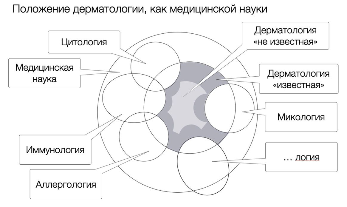
The conceptual basis of the theory


This chapter substantiates the approach to the analysis of experimental results and the construction of the conceptual foundations of the theory explaining skin conditions at the level of cell subpopulations. The basic assumptions, axioms, postulates, fundamental laws and principles are reviewed. On this basis, possibilities and practical necessity of applying methods of conceptual analysis and design for solving theoretical problems of dermatology which have technologically risen to investigations of phenomena at the level of human skin cells are shown. Motives for appealing to conceptual methods are given, and the demonstrated approach to conceptualization of a number of subject areas, opens perspectives for development of dermatology.

Measurement usually refers to the process of finding the ratio of a given quantity to another quantity taken as a unit of measurement. In turn, the results of measurement expressed by means of numbers can be subjected to mathematical processing.

"In the event of an argument," wrote the German mathematician and philosopher Leibniz, "the two philosophers will no longer have to resort to an argument, just as counting men do not resort to it. Instead of arguing, they will take pens in their hands, sit down at the boards, and say to each other, 'let us calculate.'"

By applying "more" or "less" relationships, using comparative quantitative values, it is possible to show the dynamics of change over time, to reflect the course of the disease and/or the effectiveness of the therapy used.

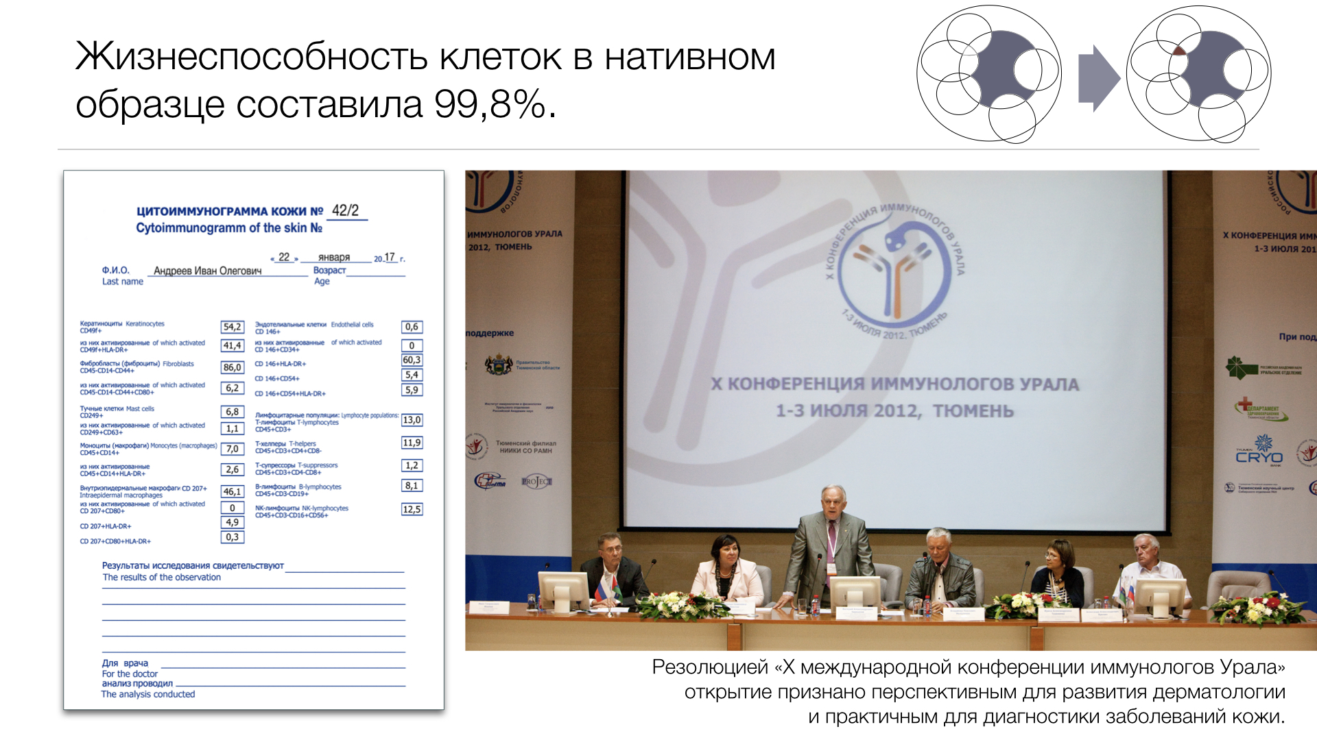
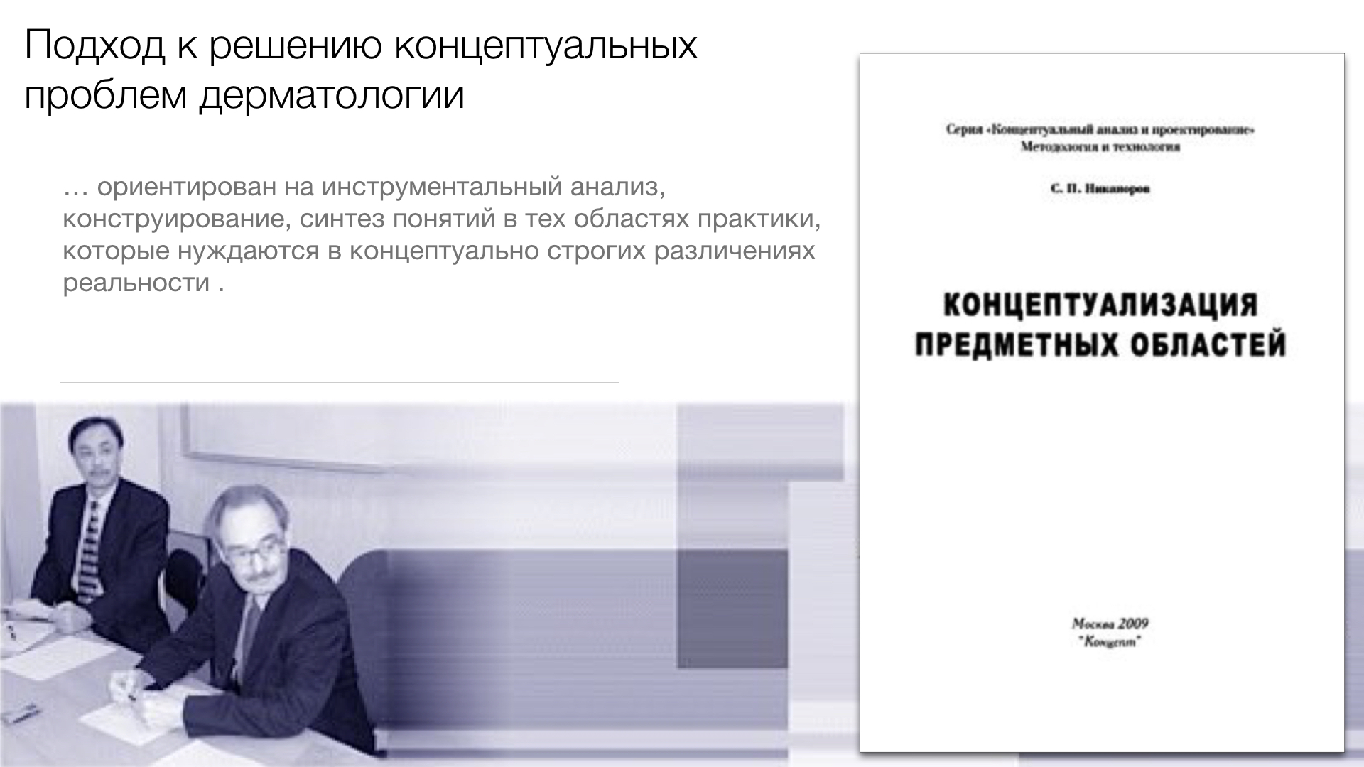
The possibility to obtain skin cytoimmunograms makes it possible to study the phenomenology of the dynamics of cell subpopulation states by conceptual methods. This implies the formation of generic and species concepts of the opened subject area based on the idea of subpopulations as specific structures of heterogeneous sets, as Nikanorov S.P. wrote about it. 

New questions arise in the practice of dermatology in connection with the opened possibilities of skin research:

  • How to carry out individual diagnosis of skin diseases, speeding up the process of treatment, if the distinctions between phenotypes of cell subpopulations are only empirical in nature?
  • How to accelerate the theoretical investigation of skin diseases ahead of practice, if problem statements and associated results are based only on a large body of empirical data from patient examinations and are therefore dependent on the development of instrumental tools for disease investigation?
  • How can we use the new possibilities of skin conditions research to raise the level of development of dermatology, which still relies on traditional methods, mostly symptomatic in nature?
  • Answers to these and similar questions lead to an understanding of a number of theoretical problems in modern dermatology:

There is a lack of an exhaustive understanding of the full diversity of cell subpopulations whose condition may influence the development of skin diseases. Reliance on empirical phenomenology is important, but extremely inert;
There is a lack of insight into the diversity of possible phenotypes of cell subpopulations. The notion of this diversity and the distinction of all possible phenotypes could lead to a wide range of new research tasks, which science is developing;
The relationships between skin cell states and pathologies for the diversity of possible phenotypes of cell subpopulations have not been established.
A solution to these problems may follow in a conceptual analysis of the phenomenology of the dynamics of skin cell subpopulation states.

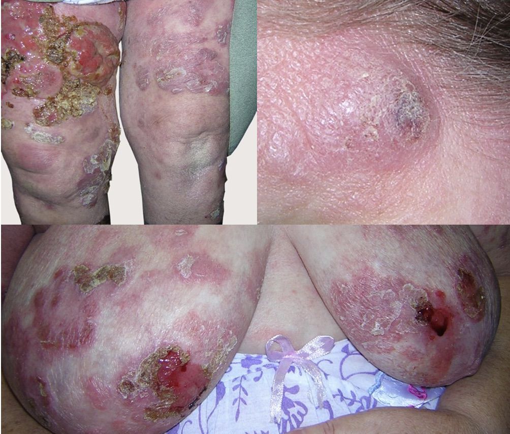
When discussing the visible part of manifestations of inflammatory processes in the skin and pathogenetic mechanisms underlying their occurrence and realized at the cell level, it should be said that they are considered as parts of a single whole. Interacting with each other, these parts determine the entire variety of rash elements and their transformations observed by the doctor.

It is this understanding that we have constantly encountered in the course of all the previous material, but not explicitly, but even as if opposing one phenomenon to another. This is not so! In practice, we are just dealing with a set of externally observed and internally invisible information about the skin cells' functions and their interrelation and interactions within the whole. More precisely, we deal with a system of heterogeneous objects united into one whole - the human skin.

However, it should be taken into account that each part of the system - in our case the cell - can be considered as an integrity consisting of its own parts, and therefore can also be studied as a system. But, in our case, it is the holistic consideration, establishment of interaction of cells (system elements) and their phenotypes in the aggregate that should be considered. 

Using the opened possibilities of skin cell subpopulation research, I offer the reader an approach to solving conceptual problems of dermatology. To emphasize the determining role of this approach in theory formation, I took the liberty of calling it the conceptual basis of the theory or the fundamental theoretical scheme.

The introduction of such an unfamiliar view of problem solving was done intentionally, in favor of maximum objectivity of judgment. The results of such an approach, which allows a deeper, more complete and more precise cognition of the properties and regularities of the object under study, are achieved by creating conceptual schemes, which are a representation of the existing material world. The essence of the approach is as follows.

1. We have chosen technology which operates with concepts based on rigorous mathematical methods and allows to deduce logically consistent consequences from the experience's statements and assumptions as the main tool of analysis of experimental data on the states and dynamics of cellular subpopulations. The grounds for the choice of conceptual methods of analysis in relation to the discovered problem are as follows:

Concepts are used in the inextricable relationship of their two essential facets: conceptual volume and content. And since the cellular level of research of skin conditions directly leads to the research of structures of subpopulations of cells formed from various sets, it gives a chance through the research of these sets as volumes of the concepts behind them to come to the content of these concepts and conceptually distinguish all variety of skin conditions;

The basic logic of conceptual methods is the logic of ascending from the observed concrete to the mentally abstract and through it to the generation of the variety of the mentally concrete. This means that a variety of all theoretically possible cellular phenomena can be derived from the limited composition of experimentally obtained structures of cell subpopulations, which will make it possible to reach a theoretically complete picture of skin states. This possibility of conceptualizing experimental results opens up the space for scientific research that is significantly ahead of the rate of new dermatological diseases;

All operations on the concepts are performed instrumentally in the conceptual technology. For this purpose, logico-mathematical tools are used: the calculus of statements, the mathematical theory of genera of structures and the apparatus of stages of sets. This allows methodically and strictly to deduce all logical consequences from the results of synthesis of concepts. Such corollaries are new concepts which were not obvious at the beginning of conceptualization and could not appear in the course of the set experiments. The ability to instrumentally generate new concepts will greatly enhance the performance of experimental dermatology by inferring from experiments corollaries that go far beyond observation.

2. It is proposed to conceptualize experimental phenomena in three subject areas constructed in the logic of deepening the distinction of skin states:

Phenomenology of static skin states. Within the boundaries of this subject area, concepts defining the full diversity of cell subpopulations, the diversity of subpopulations with the distinction of cell trait diversity, cell activity states, cell functions in the structure of a particular skin puncture biopsy, subpopulation function structures and other phenomena considered in statics can be derived;
Phenomenology of skin state transitions (state dynamics). All kinds of skin state changes can be distinguished within the boundaries of this subject area, a typology of skin state dynamics is built, a variety of chains of possible transitions, chain structures and other possible phenomena of skin state changes are established;
Phenomenology of conditioned transitions of skin states. Within the boundaries of this subject area, phenomena arising from various types of artificial and natural interventions on skin states and their dynamics can be presented; typology of interventions; typology of theoretically possible chains of transitions between skin states conditioned by interventions and other phenomena of practical influence on skin.
3. A research strategy of sequential mastery of subject areas and areas that can be synthesized from them has been chosen. This decision is due to two circumstances. First, the results of conceptualizing one, simpler subject area will inevitably influence the initial representations necessary to comprehend another, more complex one. Second, the experimental branch of dermatology, focused on the study of skin properties at the cellular level, will develop in parallel with the work with the subject areas. 

It is supposed that conceptual clearing of the phenomenology of states of skin cell subpopulations and their dynamics will allow to set tasks for investigation of medical and biological regularities between various phenomena, i.e. will lead to the very "mechanisms" of a new type of dermatology.

The results of measurements expressed in numbers can be subjected to mathematical processing and thus the phenomenology of states of cell subpopulations can be investigated by conceptual methods. It presupposes the formation of generic and species notions of the opened subject domain on the basis of idea about subpopulations as about specific structures of heterogeneous sets. The conceptual analysis of the phenomenology of skin cell subpopulation states will make it possible to set tasks for the study of medical and biological regularities between various phenomena.

There is an assumption that the use of instruments enhancing the reliability of observations can completely eliminate, if not errors, then subjectivism in the process of observation. However, this is not entirely true, because the data recorded by even the most high-tech devices does not say anything by itself. They require an interpretation based on appropriate theoretical ideas about the use of the device.

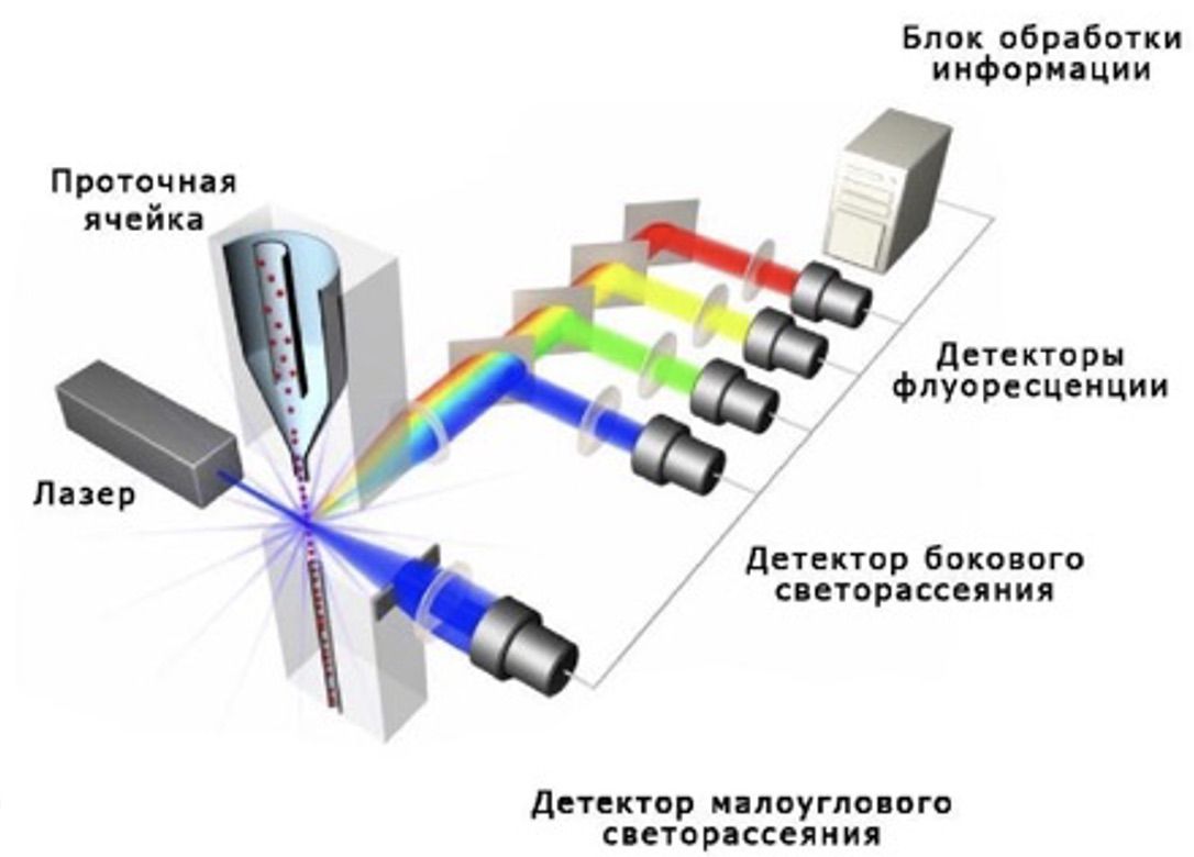
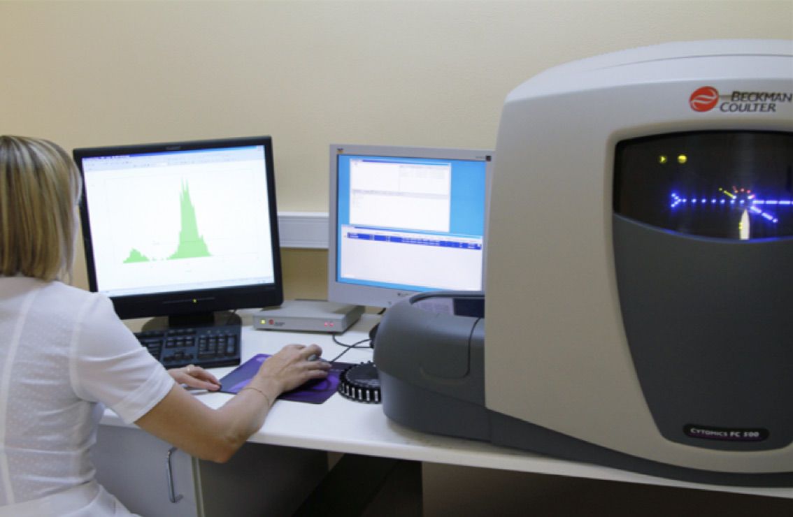
The activity of molecules on the surface of skin cells cannot be directly observed, but it can be inferred from flow cytometer readings and can therefore be considered as observable quantities.

The absence of an obvious connection between empirical (observable) and theoretical (speculative) concepts does not exclude the possibility of establishing both a logical connection and a difference between them. Developing this thought, it should be clarified that any removable observations by themselves, without their theoretical interpretation, cannot serve as a basis for conclusions or confirmation/disproof of a hypothesis or theory.

Without a theoretical explanation, findings can remain incidental and incomprehensible data, much less serve as sources of information to clarify the clinical picture of the disease observed by the doctor.

Since the data recorded by the device are still abstractions from the observed phenomena and cannot be fully correlated to the elements of the rash. Therefore, the theoretically traceable connection, is still far from true, but can be established by empirical interpretation with the clinical picture. One way or another, but empirical and theoretical concepts are most closely related, because the latter are based on observational experience, and therefore in their own way - secondary.

In the further comprehension of new knowledge, these concepts condition and complement each other, forming a perimeter of coincidence and recognition of observed pictures of the real world with descriptions obtained from the use of high-precision instruments. However, already at the empirical stage of cognition, diagnoses are introduced into dermatology with precise justifications based on observed phenomena. But they denote nothing more than the totality of the perceived picture of the world by our senses.

The transition from empirical to abstract, theoretical concepts represents a dialectical leap from the sensory-empirical to the rational-theoretical stage of research.

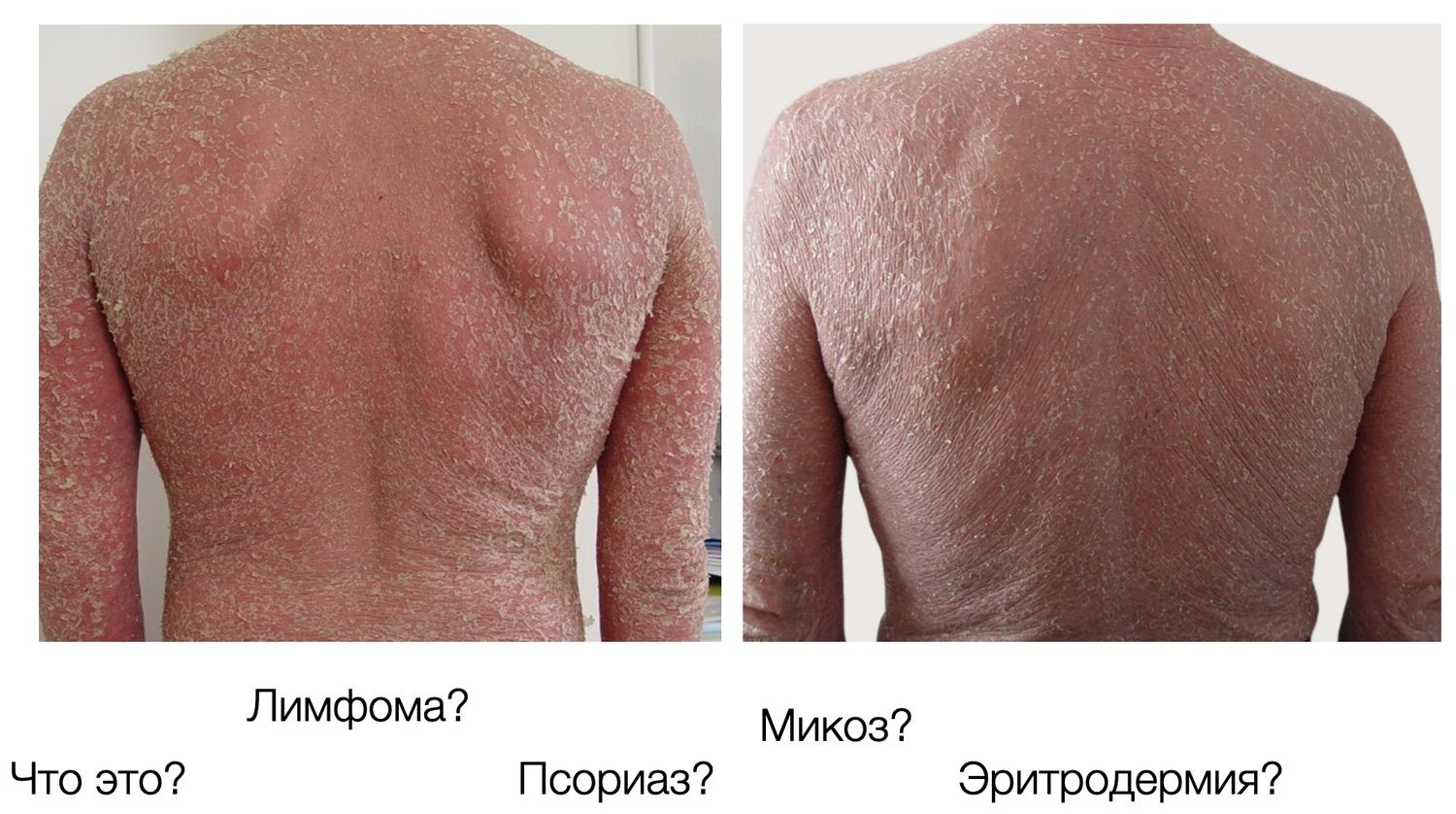
Thus, the genetically determined process of formation of a variety of specialized cell phenotypes reflecting their function is the result of coordinated expression of a certain set of genes, as a result of which, cell differentiation changing their functions, morphology and metabolic activity creates prerequisites for their high specialization and selective action, which in the end is realized in a huge variety of clinical phenomena of the same skin diseases seen by dermatologists.

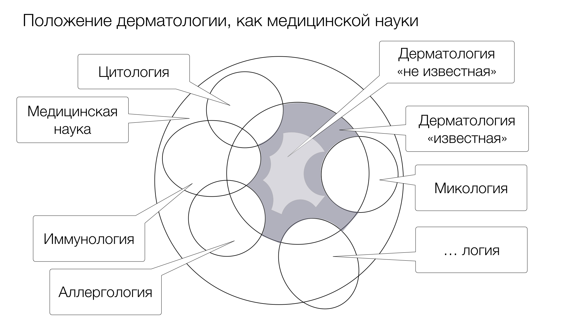
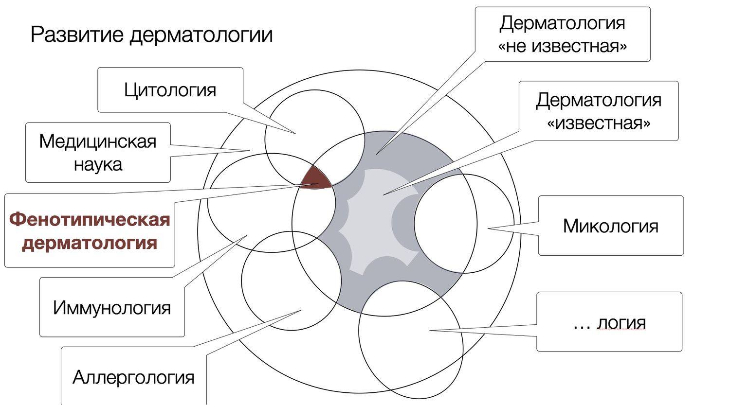
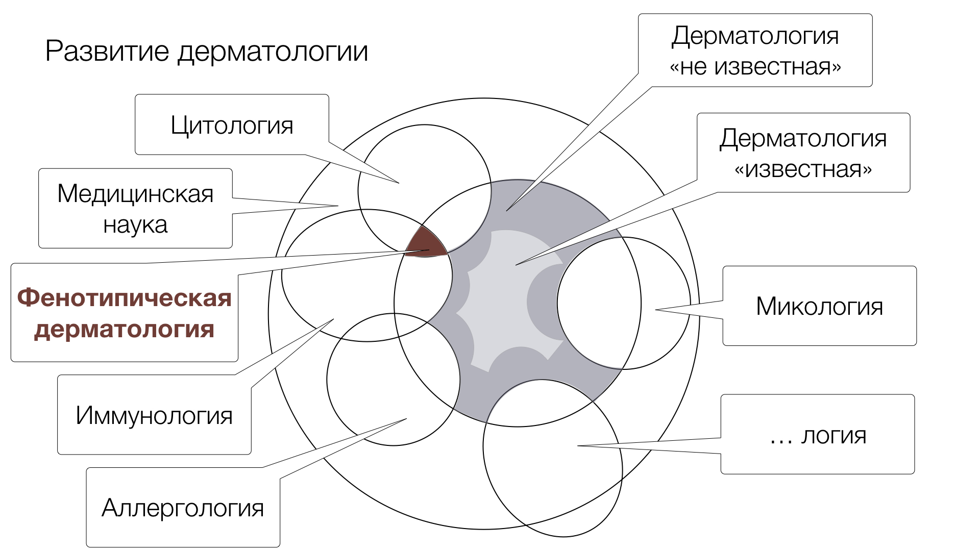
The deepening of these conceptual distinctions allows us to reach a broad class of research tasks in dermatology that relies on distinguishing phenotypes of subpopulations of skin cells - phenotypic dermatology.

 The results of conceptualization point to the possibility of a transformation of modern dermatology. A sign of this transformation is that the "unit" of the study of skin conditions becomes not the picture of its pathological symptoms, but the peculiarities of the cell subpopulation of its specific area. Each specific subpopulation, consisting of cells of all species simultaneously, reflecting specific functional-cell structure of an elementary skin fragment, represents its specific phenotype.

The transition from the symptomatic approach in the study and treatment of skin diseases to the phenotypic approach is a transition to a different, new type of dermatology - phenotypic dermatology. It should be based on the mechanisms of cytoimmunology, which is able to study and use the properties of all possible phenotypes of skin cell subpopulations.

Dermatology, as a science, is interested in objective facts that can be controlled and counter-verified, not in emotional impressions based on empirical experience, which are the domain of the subject and only. I dare say that phenotypic dermatology is the first theory describing skin functions under conditions of normality and pathology, based on a unique method and expressed in schemes of conceptual approach.

The role of theoretical assumptions is that with their help the systematization of scientific knowledge is achieved, involving the use of theoretical assertions, which, having identified and being guided by the rules of logic, can be derived from them all other assertions, including those that allow for empirical interpretation. The methodological function of theoretical concepts is related to their application to generalizations and the expansion of scientific knowledge. After all, we know that empirical generalizations reveal a certain regularity in the functioning of objects and phenomena, which in life is often beyond observation. Nor do they explain the mechanism or cause of this regularity.

Let us say that a particular diagnosis is formed by the recurrence of its symptoms and, as a consequence, the effectiveness of the remedies applied. However, if we know what mechanism predetermines the appearance of these symptoms, based on observations, but already at the level of molecules, then further on, we do not need to use each time laborious and often expensive diagnostic methods, because we know the mechanism of appearance of these or those elements of the rash. Once upon a time, an ordinary magnifying glass was a device not used by all dermatologists of the past due to the high cost of the latter. But, the knowledge obtained with it was then reflected in books, in the form of descriptions of the subtleties of distinguishing a rash, providing dermatologists with new knowledge of the clinical picture already realized in text and new concepts.

In this regard, establishing and proving the existence of links between theoretical knowledge and empirically verifiable corollaries will play a crucial role in theory. Only abstract concepts constitute the conceptual core of a theory and can explain empirical facts. Therefore, the starting point for the construction of a theory oriented toward the pragmatic realization of its conclusions should be the proposal of abstract concepts. 

Despite the seeming pragmatism of theory, only theory can be unafraid to dare to answer the questions: what, why, and how? By answering these, dermatologists of the future will be as effective as possible, both in their predictions and in the practice-oriented nature of their judgments, prescriptions, and, of course, scientific research.

Phenotypic dermatology is the language in which dermatologists must speak to each other and to dermatologists of future generations, in order to avoid losing the generational gap. It is a kind of knowledge baton with a stick of dermatological thinking.

Here we should clearly define those few but important concepts that will be taken as basic, conveying meaning. Phenotype, translated from Ancient Greek meaning "specimen," is a set of traits and characteristics inherent in a biological object at a certain stage of development. The phenotype of the object (a cell in our case), which is formed on the basis of the genotype in the process of phenogenesis, changes under the influence of environmental factors. The term phenotype was suggested by the Danish scientist, professor of the Institute of Plant Physiology of Copenhagen University, member of the Swedish Academy of Sciences Wilhelm Ludvig Johansen in 1909, in his work "Elements of the Exact Teaching of Heredity", introduced the terms: "gene", "genotype" and "phenotype", to distinguish heredity of the organism from what results from its realization.

In 1903, in his work "On inheritance in populations and pure lines," he also introduced the term "population," Latin for population, which is a set of biological entities of one species, long occupying a certain area in space and partially or completely isolated from representatives of other similar groups. In cytology, a cell population is a group of cells homogeneous according to a certain criterion. Hence, we will call a subpopulation a subspecies of cells distinguished on the basis of the function they perform.

Moving in this logic, it will not be superfluous to note that the skin as an organ will be a general population (in another word - generic) of all skin cells, in relation to which it is supposed to draw conclusions. It is worth remembering that conceptualization is the process of translating ordinary, generally accepted representations of whatever into the form of products of conceptual thinking - concepts - notions characterizing the limit of meaning distinctions.

And, a refined definition might sound as follows.  Phenotypic dermatology is a branch of theoretical dermatology that studies the phenotypic diversity of human skin cells.

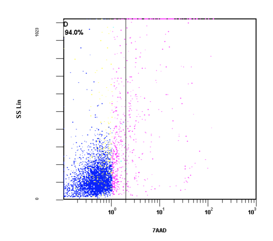
Among the most important confirmations of even the most accurate theoretical inferences is first and foremost the testing of theory in practice, it is the most significant indicator of the pragmatic nature of the research carried out. And, to establish a connection between invisible quantitative changes at the cell level and visible changes at the skin level, as well as to determine a critical value of flow cytometry parameters, is not possible from theoretical considerations. Therefore, there is a need for specific studies by means of practical observation.

Simultaneously with the received set of problems of conceptual value have opened. These are problems in relation to which it is not yet clear how to think about them in order to solve them. In order to answer these questions, one must know the whole, which Aristotle explained as having nothing to add to the existing. In other words, one must keep the whole in mind in order to be able to understand the particular in detail.

Dermatologists increasingly see the devaluation of terms in the context of progress. Borrowing terms from the literature, we then apply them to our activities (significantly changed from what they were at the time the term was described) and see that the term no longer explains what is observed and, at a minimum, we are disappointed in the descriptive properties of the term, and, at a maximum, we are on the path of a false description of the object. 

Without creating new terms and meanings, we will not be able to adequately describe the observed picture and, as a consequence, guarantee quality care for patients with skin diseases.

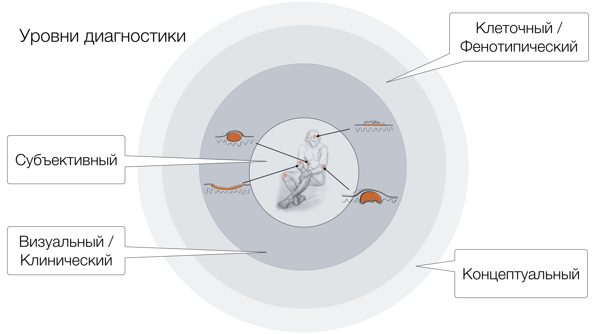
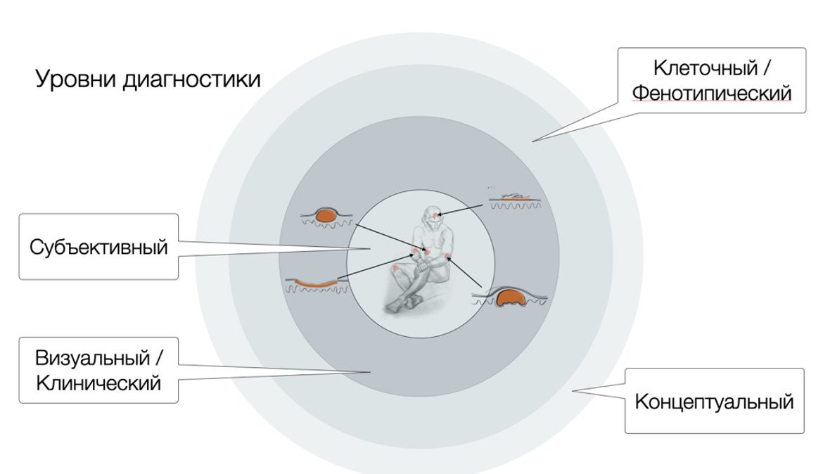
Any situation of thought activity is a division of levels of cognitive activity. In our case these are the levels of symptom reading:

  • subjective (based on the patient's assessment of his or her experiences with the ailment),
  • visual-clinical (doctor's, what I see is what I describe in familiar terms)
  • cellular (information-objective and based on unique information readable by the device and interpreted by the proposed method)
  • conceptual (reflecting the information about the patient's condition in concepts).


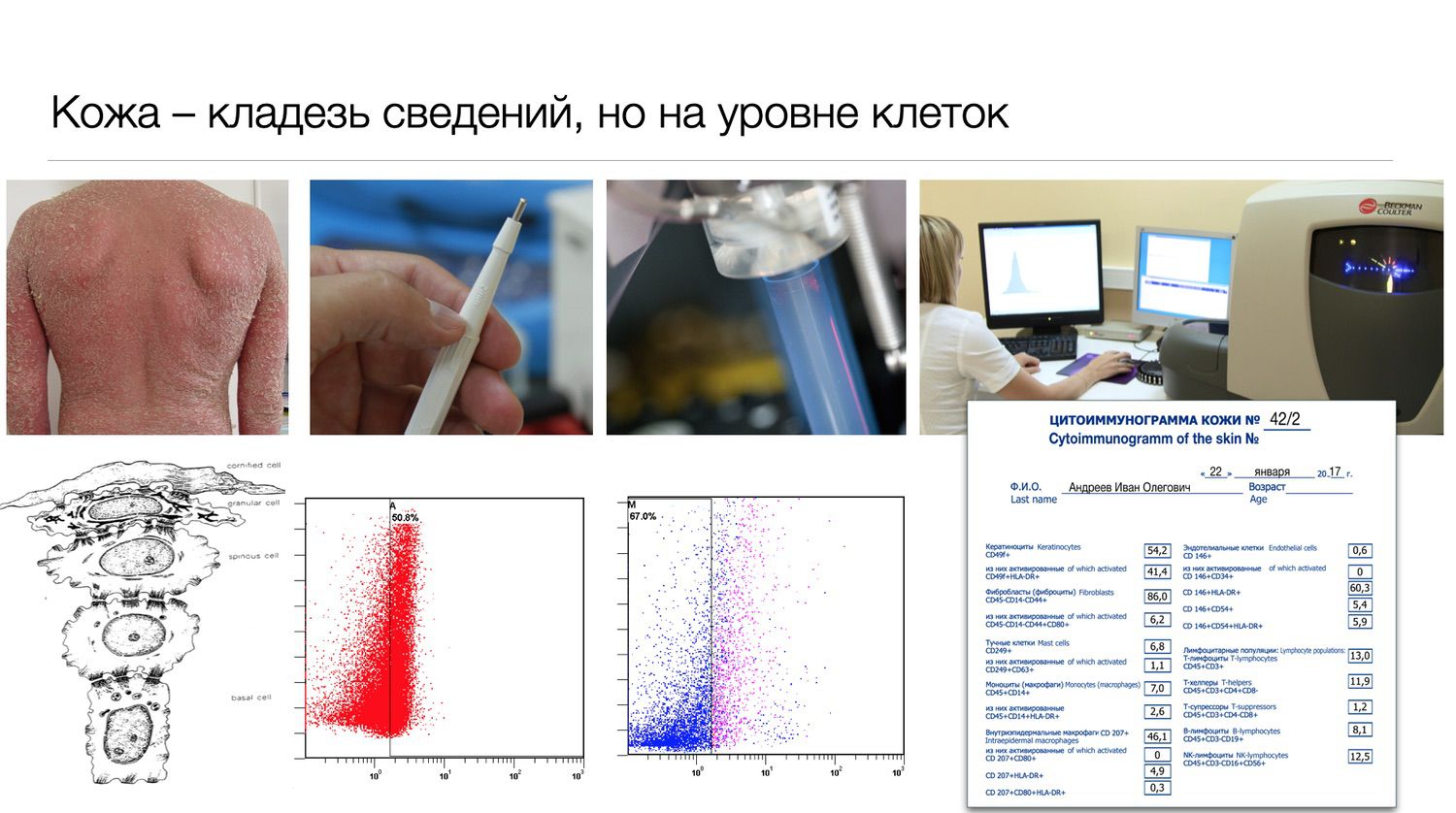
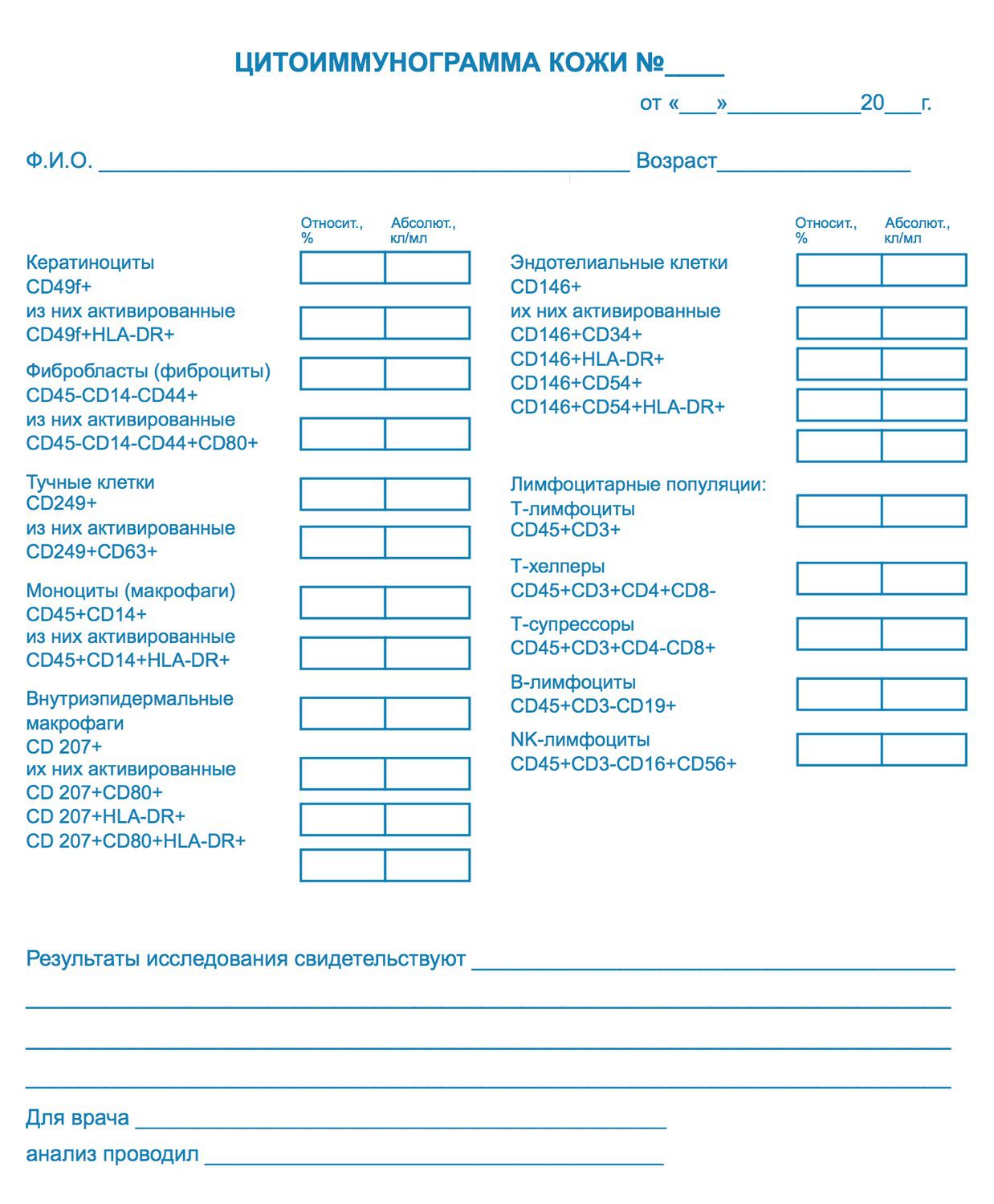
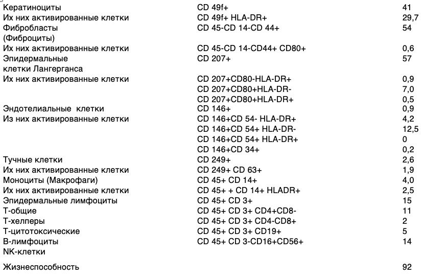
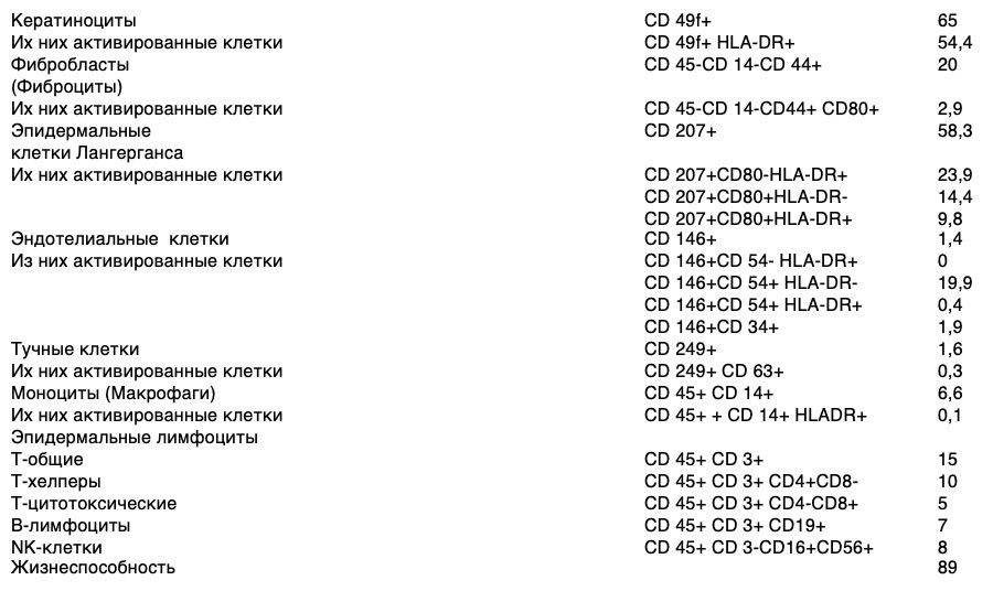
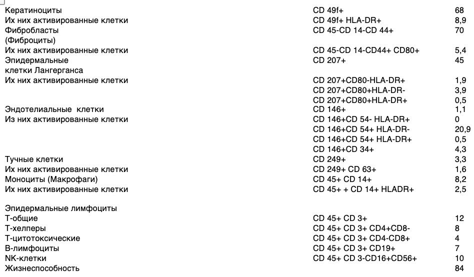
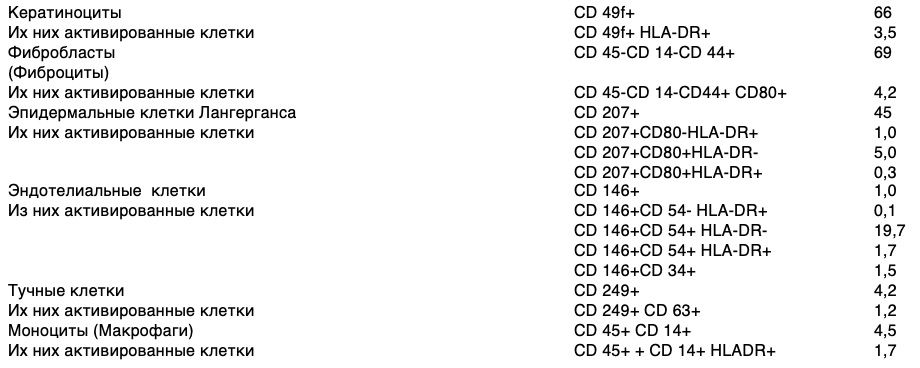
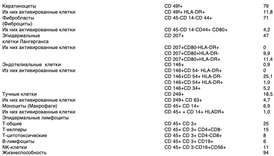
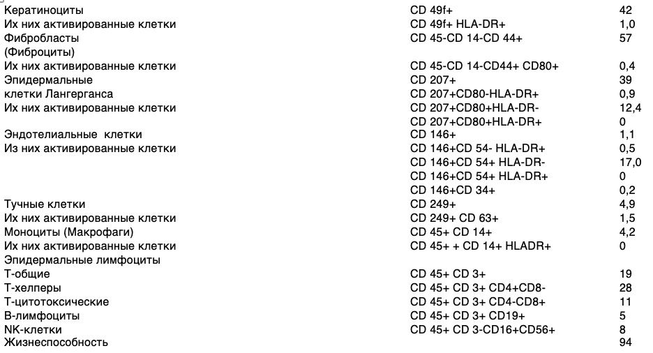
For construction of a number of concepts in the mathematical apparatus of generic structures, which is involved in the conceptual methods, adopted the initial provisions derived from the results of obtained skin cytoimmunograms. In these structures, the numbers of elements express the volumes of concepts, and the structures themselves express their contents. The representations of skin properties can be summarized in the following statements:

  • A limited variety of cell types, twelve of them (K=12), are involved in creating the phenomena of skin states. And five of them are of the same type (lymphocytes);
  • A cell of each species possesses a certain composition of traits, which are detected by markers - clusters of differentiation molecules on the cell surface (CD). Each marker acts as an unambiguous indicator of one or another manifested cell trait;
  • As science advances, the number of CDs increases and their indicative component is refined. However, a limited composition of markers is applied in each research act. Of course, this affects the accuracy of recognition of the variety of cell traits and the variety of cell types;
  • The number of potentially possible functions of each cell is determined by combinations of all its traits;
  • The number of traits taken for study is limited - twenty four for each cell of each species. The number of characteristics taken for examination is limited to twenty four, and one of these characteristics characterizes a particular kind of cell;
  • The living cells are always active. But their activity is differently manifested and influences on skin condition. The activity of a cell is a manifestation of its particular function in the structure of a particular puncture biopsy of the skin. If a cell does not show any function, we can say that its activity is zero, although it is alive. It is generally accepted that in this case the cell is passive;
  • The cells of each species may exhibit (or not exhibit) one or several different functions at once. This is determined by the composition (combination) of the cell traits;
  • The number of potentially possible functions of each cell is determined by all combinations of all its traits;
  • Cells are combined into subpopulations. Each subpopulation involves a specific fraction of cells of each species with its different functions. All theoretically conceivable subpopulations are admissible, since there are no restrictions on their diversity or they are not known;
  • A subpopulation with a specific composition of cells, each with a specific composition of functions, forms a subpopulation phenotype;
  • Each combination of subpopulation phenotypes uniquely corresponds to a specific immunological skin condition expressed in a clinical or physical symptom.

These statements serve as the basis for the generation of concepts, each of which defines a class of possible phenomena and allows for the differentiation of the variety of situations that arise in the skin when considering its findings at the cellular level. In what follows, with brief explanations, only those concepts that define static skin phenomena are presented. They can be regarded as instantaneous "slices" of its state dynamics. Concepts are given in the mathematical apparatus of genera of structures, which is involved in conceptual methods.

The concept "Variety of Skin Cell Species Functions".


Since any cell of any species can exhibit any combination of features, this structure establishes a complete diversity of all combinations of features that only a skin cell of any species can have. Given that each combination of features of a cell indicates the activity of its particular function, this structure simultaneously expresses the full diversity of functions that cells can activate, regardless of their species.

If the number of features of each cell is equal to P, and one of them is used to determine the type of the cell itself, the variety of combinations of features and, consequently, the functions of cells can reach the number 2 (P-1)-1. Here it is taken into account that if a cell does not exhibit any features (empty subset of features - ∅), it is functionally passive. Taking into account the known number of features of a cell of each species (P = 24) it turns out that the maximal power of hypothetical variety of functions of one cell of any species (F) is more than 8 million (specifically: F = 8 388 607). And each of these functions can be distinguished.

In order to demonstrate possibilities of the method let us take a basic set:

X1 is the set of features of a cell of any species that are experimentally detected by clusters of differentiation.

The genus relation will be expressed by the following structure:

D1 ∈ B(X1).

The type of this structure: "The set of all subsets of the cell's traits".

Example.

Suppose a cell does not have 24 features, but only 3. That is, let the set X1 = {a, b, c}.

Then combinations of these features are formed as follows:

B(X1) = {{a}, {b}, {c}, {a,b}, {a,c}, {b,c}, {a,b,c}, {}}.

It turns out that the number of combinations is 23 (two to the power of 3). If we ignore the empty set, which indicates a passive cell, then the number of "active" combinations is 23-1.

Each combination is a function of the cell. Hence, seven combinations are formed from three features - seven functions:

{a}, {b}, {c}, {a,b}, {a,c}, {b,c}, {a,b,c}

This is the first result of conceptualization. It allows to theoretically define each function of a cell, investigating its influence on the state of health; to set tasks to find effective ways to detect functions; to look for ways to activate each function or to suppress it. Currently, of all the variety of skin cell functions, a tiny fraction has been studied dermatologically. It is possible that some potential functions will never manifest or will manifest in some unique situations. But, in the absence of substantiated statements to that effect, the occurrence of unfamiliar functional skin cell phenomena cannot be ruled out. 

The concept "Species Cells with Functions".


Let's try to extend the conceptualization by taking not only the features of cells of one species, but a multitude of cell types. The basic sets will be as follows:

X1 - the set of cell features of any species that are experimentally detected by special markers;

X2 - the set of skin cell species. These are all those distinct cells that are involved in the creation of skin health phenomena.

The genus relation has the following form:

D2 ∈ X2 × IN(X1). Here "×" is the sign of the Cartesian product of sets.

The type of this structure is: "The set of cell types and the set of their functions".

This structure defines all situations in which it is necessary to distinguish specific species cells with all their functions. The variety of such distinguished combinations is over 100 million (specifically: F = 100,663,284).

This concept is useful for setting and solving narrow theoretical problems in dermatology. Thus, consideration of cells with their full set of functions allows to investigate the peculiarities of action of all functions of each species cell. The question can be answered whether the fact that the same function is manifested in different species cells at the same time will change the effect on the skin. Assuming that the number of species cells is K = 12, and the number of functions of each species cell is F = 8,388,607, then the variety of "species cells with their functions" compositions (S) is S = K × F = 100,663,284. Of course, it will take many years to empirically study the properties of all these situations. But the room for theoretical research is open.

Example.

Suppose we have only three cells. That is to say

Х2 = {1. 2. 3}.

And each cell (from the previous example) has 7 functions:

X1 = {{a}, {b}, {c}, {a,b}, {a,c}, {b,c}, {a,b,c}}.

Then the number of "situations" where three cells can act with different sets of their functions is determined by the direct product:

X2 × X1 = 1{a}, 1{b}, 1{c}, 1{a,b}, 1{a,c}, 1{b,c}, 1{a,b,c}, 2{a}, 2{b}, 2{c}, 2{a,b}, 2{a,c}, 2{b,c}, 2{a,b,c}, 3{a}, 3{b}, 3{c}, 3{a,b}, 3{a,c}, 3{b,c}, 3{a,b,c}.

However, the real situations in which a particular panch biopsy of the skin reveals different numbers of species cell compositions are not yet expressed here. In this sense, a concept with a different generic structure should be recognized as more constructive and meaningfully rich.

 Here "×" is the sign of the Cartesian product of sets. 

The concept "Variety of species cells with sets of their functions"


The basis sets of this concept will be as follows:

X2 - the set of skin cell species;

X3 - the set of cell functions.

The generic relation has the following form:

D3 ∈ IN(X2 × IN(X3)).

The type of this structure is: "The set of subsets of species cells together with the set of subsets of their functions".

It expresses all situations where, in a particular skin puncture biopsy, it is possible to consider separately such subpopulations in which cells of all species (or only a few) are collected together with their manifested functions. Their diversity is large but measurable, specifically it is 2100 663 284.

Such situations resulting from the evolution of skin cell states are "subpopulation phenotypes". They are an important diagnostic sign to judge the course of processes in the skin. Phenotype should be understood as a set of functionally significant markers characteristic of certain stages of differentiation, proliferation, activation or apoptosis. The relative and absolute number of cells having one or another phenotype is precisely the end result of cellular states. The diversity of them is great:

Single cell types and their functions. These are all those phenotypes of subpopulations that are determined by the previous genus of the structure;
Phenotypes of subpopulations formed from several cell types (not all) at one time together with their different functions;
Phenotypes of subpopulations formed by cells of all species, with each cell species exhibiting one or two of its functions;
Phenotypes of subpopulations formed by cells of all species, with each cell species exhibiting all its functions simultaneously.
This concept opens up the possibility to distinguish phenotypes with complete/incomplete sets of cell species with all different activities of functions, including those in which all cell species exhibit all their functions simultaneously. Based on it, new classes of problems can already be set to investigate health/disease situations with different compositions of functions in complete sets of cell species and other situations. But these conceptual constructions do not yet take into account the circumstances when in a skin biopsy a certain proportion of cells of each species with specific (different) functions turns out. It will be another concept that expresses the full diversity of phenotypes of subpopulations of cells. At the same time, unusual theoretically possible situations can be expressed. For example, when there is a complete diversity of cells of all species, there will be cells with different sets of functions among cells of the same species. This will force researchers to answer a series of questions about what are the properties of such phenotypes of cell subpopulations and how does this relate to the pathologies that are the subject of dermatology as a science of skin and its diseases.

Example.

Suppose we have only cells of three kinds. That is to say.

Х2 = {1. 2. 3}.

And each cell (from the previous example) has 3 functions:

X3 = {m, n, o}.

Then we can see (distinguish) the following situations:

Situation 1: 1m,

Situation 2: 1n,

Situation 3: 1o,

Situation 4: 2m,

Situation 5: 2n,

Situation 6: 2o,

Situation 7: 3m,

Situation 8: 3n,

Situation 9: 3o,

Situation 10: 1n, 1o,

Situation 11: 1m, 2m,

Situation 12: 1m, 2n, 3o,

Situation ...: 1m, 2m, 2n, 3n, 3o,

Situation ...: 1m, 1n, 1o, 2m, 2n, 2o, 3m, 3n, 3o.

The results of the performed conceptualization indicate that the "unit" of the study of skin conditions becomes not the picture of its pathological symptoms, but the features of the cell subpopulation of its specific area. Each subpopulation, consisting of cells of all species simultaneously, reflects a specific functional-cell structure of the elementary skin fragment and represents its specific phenotype.

The conceptualization of the phenomenology of skin cell subpopulation states and its dynamics reveals classes of potential situations, which are still indistinguishable in dermatology. This circumstance explains the fact that the gigantic variety of skin diseases that practitioners deal with have not yet acquired their own names, much less unique treatment approaches.

Further deepening of conceptual distinctions of states of skin cell subpopulations and its dynamics, deployment of generic structures of these dynamics will allow to bring hitherto latent circumstances of skin pathologies to the explicit level. These possibilities give an expansive character to the generation of tasks of theoretical research, which cannot be set in any other way. Thus, the derived tasks become the basis for setting new, increasingly "subtle" experiments and the generator of requirements for the development of the experimental base of dermatology.

The rigor of conceptual distinctions allows us to reach a broad class of research tasks in dermatology based on differences in phenotypes of skin cell subpopulations, and the transition from the symptomatic approach to treatment of skin diseases to the phenotypic approach is a transition to dermatology of a new type - phenotypic dermatology. It should be based on information about the properties of all possible phenotypes of skin cell subpopulations. Phenotypic dermatology, which describes the functions of skin cells under normal and pathological conditions, based on the way cytoimmunograms are obtained and expressed in conceptual approach schemes, is interested in objective facts that are possible to control and counter check.

A hypothetical solution to a scientific problem


Translated from the ancient Greek, the term "problem" means a difficulty or obstacle, to overcome which practical or theoretical efforts are made. Thus, in order to confirm the scientific hypothesis and to achieve the goal, the work set a number of the following tasks, which were solved in the course of the study:

To analyze the current state of dermatology and substantiate the problems that arise with the appearance of skin cytoimmunograms.
To substantiate the conceptual "unit" of diversity of skin conditions at the cellular level.
Determine a phenotype of human skin cell subpopulations.
Develop methods of obtaining a viable heterogeneous skin cell population, determining skin cell subpopulation composition and obtaining skin cytoimmunograms.
Substantiate an approach to building a field of concepts, defining the full diversity of skin cell subpopulations for advanced scientific research on human skin cells and develop these concepts.
Experimentally demonstrate the practical utility of using flow cytometry and constructed concepts in the study of properties of skin cell subpopulations and their application in diagnostic and therapeutic practice.
Develop criteria for application of flow cytometry for diagnostics in dermatology.
To develop a diagnostic panel for the study of individual subpopulations of skin cells.
As an example of practical utility develop a device for activation of reparative potentialities of skin cells.
On the basis of the methodology to develop a wound healing agent, a method of its production and a method of treatment of wounds of various etiology by the obtained remedy.
To formulate new scientific tasks for the development of methods of forecasting, diagnostics and treatment of skin diseases.
The formulated research tasks allow us to propose a scientific hypothesis for their solution, which, having received reproducible confirmation, becomes the basis for the theory. The hypothesis in this paper is that the scientific problem will be solved if:

The phenotype of skin cell subpopulations will be taken as the "unit" of the variety of skin states at the cellular level, representing their special, different from others set of cells of the elementary skin fragment, forming a specific functional structure from cells of all species simultaneously;
A theory explaining skin conditions at the level of its phenotypes will be built on the basis of a toolkit operating with concepts and suitable for deriving logically strict consequences from the results of experimental skin research at the cellular level;
The approach to theory building will be based on development and synthesis of fields of concepts for definition of skin states in statics; transitions of skin states; transitions of skin states caused by various kinds of artificial and natural interference with it at the cellular level;
A number of conceptual constructions of the emerging theory will be tested experimentally in the course of diagnostics of skin conditions at the level of phenotypes of its cell subpopulations and on this basis it is possible to obtain practical results, significant for development of therapeutic practice;
From the constructed conceptual space of the theory, new classes of problems will be derived, developing dermatology at a rate outpacing the speed of the complication front of skin pathology.
The intermediate position between scientific problems and problems is occupied by problems that involve a choice between alternative possibilities for their solution.

A scientific hypothesis is not the ultimate goal of research. On the contrary, it represents the beginning of the path to pattern discovery, since it is the thoroughly tested, confirmed, and substantiated hypothesis that becomes a theory.

However, there is hardly a credible logical transition from empirical facts to theoretical laws, since there are no theoretical concepts in empirical cognition. The only way to discover them is by proposing hypotheses that are general in form and profound in content, the consequences of which are reliably confirmed by observation or experiment.

Thus, it is planned to solve the scientific problem of building a theory explaining skin conditions at the level of phenotypes of its cell subpopulations, suitable for practical and theoretical development of dermatology and pragmatic use of knowledge about the consequences of dynamics of phenotypic diversity of human skin cells. 

Provisions of theory


Theories must not only explain the situation as it is, our old experience must also be deduced from them.

Karl Popper

In the statements presented, theory brings isolated facts about the role of human skin cell phenotypes into a logical relationship, and most importantly, seeks to deduce them by deductive rules from the general premises that serve as empirical and theoretical knowledge. Creation of a theory as a system of scientific knowledge is a verification of a separate hypothesis on the basis of which it was possible to deduce the provisions of phenotypic dermatology:

  • A method of obtaining a viable heterogeneous population of skin cells, allows the separation of skin cells with preservation of their viability for further diagnostic studies, isolation and cultivation of individual populations, as well as the selection, testing and development of drugs for the treatment of skin diseases.
  • Technical result that includes skin biopsy sampling, tissue homogenization, filtration of homogenate, centrifugation of homogenate, determination of cell viability, incubation of cells with monoclonal antibodies conjugated with fluorochromes, skin cell phenotyping with determination of cell number of a certain phenotype - is a method for determination of skin cell subpopulation composition and method for obtaining skin cytoimmunograms.
  • Method of determination of subpopulation composition of skin cells enables to perform quantitative and functional assessment of the state of subpopulations of viable skin cells in different age and sex groups of patients and to correlate structural and functional parameters of the examined cells.
  • It is necessary to consider subpopulation phenotype as a "unit" explaining the variety of skin conditions at the cellular level. It represents a subpopulation consisting of skin cells of the same species having a distinctive (specific) functional-cell structure of an elementary skin fragment. This "unit" of research allows to distinguish subpopulations of cells with all various functional activities, including those in which all cell types simultaneously exhibit all their functions. On the basis of distinguishing the properties of specific phenotypes of subpopulations, new classes of tasks for research of health/disease situations can be set.
  • An effective tool for developing theoretical knowledge of skin conditions derived from experimental data should be one that is based on the synthesis of the methods of experimental dermatology and immunology with the methods of conceptual analysis and synthesis of complex subject areas. This synthesis allows to logically strictly form and represent for biophysical research in an explicit form the concepts of a full variety of skin states, distinguishable at the cellular level.
  • Rational approach to construction of notions about phenotypes of skin cell subpopulations determining their diversity and dynamics consists in consecutive explication of experimental results in the form of development of generic conceptual schemes of phenomenology of skin states in statics, phenomenology of skin state transitions (dynamics); phenomenology of skin state transitions caused by different types of interference and subsequent synthesis of these conceptual schemes. This approach will allow generating multiple concepts, allowing to distinguish and investigate properties of the whole variety of components, from which the real picture of skin states is formed.
  • Concepts of phenotypes of skin cell subpopulations and other phenomena related to skin conditions should be used as tasks for setting experiments on research of skin properties, conducting development on creation of diagnostic and therapeutic devices and preparations, setting new classes of research tasks. Deepening of conceptual distinctions of states of skin cell subpopulations and its dynamics in the form of unfolding of generic structures of these dynamics and interpretation of the terms of formal theory will allow to bring latent circumstances of skin pathology to the explicit level and create new means of treatment of skin diseases.
  • Developing directions of dermatology, which uses phenotypes of subpopulations of skin cells as "units" for investigation of skin conditions, can be generated not in the form of insights of talented researchers, but in the form of tasks for revealing biophysical properties of species concepts of formal theory methodically derived from constructed generic conceptual schemes. This kind of work with the unknown in dermatology will create a field of promising research, years ahead of the evolution of skin pathology.
  • The cognitive possibilities of studying phenotypes of skin cell subpopulations and specific rules of their study form a new field of medicine, which should be called phenotypic dermatology. The transition to it will enhance the intellectual power of the paradigm of diagnosing and treating skin diseases. 

In addition, phenotypic dermatology, as a theory, has several functions:

  • analyzes, classifies, and systematizes numerous facts;
  • provides the user with conceptual structures;
  • interprets and explains specific facts of reality;
  • stimulates the development of new terms and their pragmatic use;
  • allows predicting future developments and observed phenomena;
  • predicts the appearance of new phenomena.

As we know, the axiomatic method, first used by the ancient Greek mathematician Euclid, postulates that all the statements contained in a theory must be deduced logically from a small number of initial premises, accepted without proof. Obviously, such an ideal can never be achieved in theories that rely on observations, experiments, and empirical facts. New observations and experiences, as well as the synthesis of scientific knowledge, force us to reconsider previous attitudes and principles. Nevertheless, the very pursuit of such an ideal forces us to search for a logical connection between judgments that are different in their generality, so that a holistic view of the object of study is achieved and it becomes possible to foresee the future.

Scientific novelty


The scientific novelty of the performed work consists in the following:

The possibility of quantitative, morphological and functional evaluation of viable skin cells by flow cytometry was shown for the first time.
In the course of experiments for the first time cytoimmunograms of skin were obtained, allowing to investigate and display in figures the subpopulation composition of skin cells and their functional activity. The method of obtaining cytoimmunograms made it possible for the first time to give an intravital characteristic of the morphofunctional state of human skin cells within a single biopsy specimen.
A diagnostic panel of monoclonal antibody sets to different differentiation clusters that most accurately characterize the dynamics of the states of skin cell subpopulations was proposed and introduced for the first time:

  • CD49f+ keratinocytes, of which CD49f+HLA-DR+ activated;
  • fibroblasts (fibrocytes) CD45-CD14-CD44+, including activated CD45-CD14-CD44+CD80+
  • CD249+ mast cells, including activated CD249+CD63+;
  • CD45+CD14+ monocytes (macrophages), including activated CD45+CD14+HLA-DR+;
  • intraepidermal macrophages CD207+, including activated CD207+CD80+, CD207+HLA-DR+, CD207+CD80+HLA-DR+;
  • CD146+ endothelial cells, including activated CD146+CD34+, CD146+HLA-DR+, CD146+CD54+, CD146+CD54+HLA-DR+;
  • CD34+CD45dim epithelial cells;
  • lymphocyte populations: CD45+CD3+ T-lymphocytes, CD45+CD3+CD4+CD8- T-helpers, CD45+CD3+CD4-CD8+ T-suppressors, CD45+CD3-CD19+ B-lymphocytes, CD45+CD3-CD16+CD56+ NK-lymphocytes.

For the first time, it was shown that the phenotype of subpopulations should be taken as the conceptual "unit" explaining the diversity of skin conditions. A subpopulation phenotype is a special, distinct subpopulation consisting of skin cells of the same species and having a specific functional-cell structure of an elementary skin fragment. Transition from the symptomatic approach in skin research and treatment of skin diseases to the phenotypic approach is a transition to a new type of dermatology, which should be based on the mechanisms of cytoimmunology capable of investigating and using knowledge about the properties of all possible phenotypes of skin cell subpopulations.
For the first time, a toolkit for constructing representations of the diversity of phenotypes of skin cell subpopulations has been proposed. The toolkit is based on synthesis of methods of experimental dermatology and immunology, which have technologically risen to the level of studying and operating skin cell complexes, with methods of conceptual analysis of experimental results. This idea showed its productivity in generating a large number of classes of new concepts in dermatology.
For the first time, an approach to constructing notions about phenotypes of subpopulations of skin cells determining their diversity and dynamics has been developed. The approach is based on the fact that with the help of conceptual methods three subject areas are explicated, arranged in the logic of increasing complexity and at the same time deepening the distinctions of features of possible skin phenotype states:

  • Phenomenology of skin states in statics.
  • Phenomenology of skin state transitions (dynamics of states).
  • Phenomenology of conditioned transitions of skin states.

The constructed concepts of the selected subject areas have allowed to distinguish and investigate properties of the whole variety of components, from which the real picture of skin states is formed.
A number of scientific tasks of phenotypic dermatology were formulated in relation to approaches to the study of skin conditions for the development of methods of prediction, diagnosis and treatment of skin diseases.
The possibility of using flow cytometry and conceptually constructed variety of phenotypes of subpopulations of cells to improve the quality of diagnostics in dermatology and use the obtained knowledge in the processes of diagnosis and treatment of skin pathologies was experimentally confirmed.
On the basis of synthesis of experimental and conceptual developments, it seems possible to change the pattern (paradigm) of approaches to the diagnosis and treatment of skin diseases and the development of new drugs for dermatology. The near future of this shift has already manifested itself in a series of successive inventions creating a frontline of practice-oriented innovations and discoveries. 

An example of a pragmatic realization of the theory


The results of the conceptualization revealed classes of potential situations that have not yet been distinguished in dermatology and have not been the subject of study and use in practice.

For example, classical dermatological approaches to wound healing are based on various types of external therapy aimed at rapid closure of a tissue defect "at any cost", including scar formation. And since the main links of the wound evolution are interpreted in terms of latent chronic autoimmune pathology, all existing therapeutic measures are often reduced to antibacterial and immunosuppressive influences.

However, studies by several authors have established the involvement of the immune system in the regulation of the processes of physiological and reparative regeneration of tissues and organs. In this regard, it is of interest to search for ways to influence regenerative processes by influencing the natural mechanisms of the immune system through stimulation of reparative processes.

In the variety of the constructed concepts we identified separate types (terms), which could be of importance for the actual practice of skin pathologies diagnostics. Thus, in one of the conceptual schemes, a species concept of one of the skin phenotypes was highlighted, in relation to which the task of developing a technical device for selective in vitro activation of skin cells of a particular phenotype was set.

Taking into account the results of conceptualization, this task was formulated as follows:

1. Among the full diversity of phenotypes of species cell subpopulations with sets of their functions given by conceptual scheme D3, find such a phenotype which is defined by the following term:

TR1 = {d ∈ D3 I (Pr1d ∈ X2)}

2. To develop a technical device capable of activating the cell functions of this set.

The relevance of solving this problem was determined by the RF Government Decree and the Ministry of Economic Development Order on ensuring state quality control for research of perfumes and cosmetics in order to carry out conformity assessment procedures to the requirements of the technical regulations of the Customs Union "On Cosmetics Safety" (TR TS 009/2011).

The solution to this problem was the invention of a skin cell reparative potency amplifier. On the one hand, this invention is a response to the need to introduce expert systems into medical practice. On the other hand, it is the first of many possible steps toward a therapeutic tactic in which wound healing becomes a controlled process with a given outcome requirement.

His utility model relates to biotechnology in cellular medicine, namely devices for working with human skin cells, creating a practical convenience of in vitro activation and introduction of activated skin cells into the wound. This invention opens up a number of possibilities for so-called "targeted delivery" of drug substances, the prospects of which in practical dermatology have been outlined earlier by Russian and foreign authors.

Amplifier of reparative potencies is the first practical step in the direction of phenotypic dermatology, the success of which will be based on conceptually strict differentiation of properties of various phenotypes of skin cell subpopulations and targeted regulation of these properties (activation, suppression, enhancement, modification, etc.) taking into account individual characteristics of patients.

The term "amplification" introduced by R. Descartes in "Metaphysical Reflections" meant "amplification", but we use this word as a name for the technical invention, interpreting it as an activator of reparative potencies that a skin cell has and that can be functionally progressively manifested.

In the implementation of the utility model the set task is solved by achieving a technical result, which is to ensure limited contact of the active substance in the form of a gel with the activated skin cells and their localization in a separate container with increased isolation from the external environment. 

The technical result is achieved by activation of reparative potentialities of skin cells in a device containing opaque material containers in the form of truncated cones inserted into each other, which additionally have on their lateral surface protruding annular flanges with symmetrical facets for sealing, and the upper truncated cone is filled with active substance Cellgel, the upper end of the upper truncated cone is covered by a flat blind cover and its lower end is a membrane with evenly distributed holes of diame In general, the device consists (figure ) of:

an upper truncated cone having an annular shoulder on the lateral surface projecting outwardly from the cone,
an annular shoulder on the lateral surface of the upper truncated cone, projecting outwardly from the cone to secure the lower truncated cone
an annular shoulder on the upper face of the upper truncated cone, projecting outward of the cone to secure the lid
a blind flat lid,
a diaphragm in the lower end of the upper truncated cone,
evenly spaced holes of 5-7 microns in diameter in the diaphragm of the lower end of the upper truncated cone,
the inner cavity of the upper truncated cone filled with the active substance Cellgel,
the lower truncated cone,
an annular shoulder on the upper end of the lower truncated cone projecting inside the cone
the bottom end of the lower truncated cone, which is blind,
the inner cavity of the lower truncated cone, which is filled with a divided viable heterogeneous population of skin cells in the form of a suspension.
Tightness of connection of the upper truncated cone with the lower one is provided by protruding ring collars (2 and 9) on their lateral surface and due to cone shape of the vessels as a result of constant compression of the lower truncated cone to the upper one. The collars have symmetrical chamfers to facilitate connection of the truncated cones to each other. In addition, the tightness of the lower truncated cone is further ensured by a hydraulic seal formed by the active substance in the upper truncated cone, which prevents possible penetration of anything from the environment into the activated skin cells.

Provision of limited contact of activating substance, which allows not only to cultivate skin cells, but also to activate them to produce biologically active substances contributing to reparative potencies) with activated skin cells occurs as a result of placement in the upper truncated cone of the active substance, biologically active molecules of which, due to density gradient, diffuse through a membrane with hole diameter of 5-7 µm to activated skin cells located in the lower truncated cone. The diameter of holes in the membrane is chosen based on the size of skin cells, which is from 8 microns to 15 microns, to prevent penetration of skin cells from the lower truncated cone into the activating substance. Thus, a portion of the activating agent (7) is immersed in a suspension of a divided viable heterogeneous skin cell population (11). The upper truncated cone is provided with a blind flat lid (4) to ensure tightness.

Provision of limited contact of activating substance, which allows not only to cultivate skin cells, but also to activate them to produce biologically active substances contributing to reparative potencies) with activated skin cells occurs as a result of placement in the upper truncated cone of active substance, which biologically active molecules, due to density gradient, diffuse through a membrane with a hole diameter of 5-7 µm to activated skin cells located in the lower truncated cone. The diameter of holes in the membrane is chosen based on the size of skin cells, which is from 8 microns to 15 microns, to prevent penetration of skin cells from the lower truncated cone into the activating substance. Thus, a portion of the activating agent (7) is immersed in a suspension of a divided viable heterogeneous skin cell population (11). To ensure tightness, the upper truncated cone is provided with a blind flat cap (4).

All structural elements of the device are made of opaque material chemically inert with respect to the activating agent and separated viable heterogeneous skin cell population, e.g. medical polypropylene.

The device works as follows. Initially, the upper truncated cone (1) is filled with the active substance, e.g. Cellgel (7), and a blind flat cap (4) is fixed on the shoulder (3) to seal it. The lower truncated cone (8) is filled with a divided viable heterogeneous skin cell population as a suspension (11). Connect the truncated cones to each other by inserting the upper truncated cone (1) into the lower one (8), until they are fixed by collars (2 and 9). After that, biologically active molecules from the active substance Cellgel (7) start diffusing into the divided viable heterogeneous skin cell population along the density gradient through holes (6) 5-7 µm in diameter in the membrane (5) of the upper truncated cone (1), activating epidermal and dermal cells in suspension (11), which start producing biologically active substances. After the required activation time has elapsed, the shoulder (9) is unbent, the truncated cones are separated and the activated skin cells are extracted from the lower cone (8).

Thus, between the claimed technical result and essential features of the invention there is the following cause-effect relationship: increasing isolation from the external environment is provided by making the device from opaque material, preventing penetration of sunlight to the active substance and activated skin cells; increasing convenience of extracting activated skin cells is provided by localizing them in a separate container - the lower truncated cone; providing limited contact of the active substance in The diameter of holes in the membrane is chosen based on the size of skin cells, which is 8 to 15 microns, to prevent penetration of skin cells from the lower truncated cone into the active substance. The active ingredient used is Cellgel, which allows not only to cultivate skin cells, but also to activate them to produce biologically active substances contributing to the reparative potency of skin cells. Activated skin cells are a divided viable heterogeneous population of skin cells in the form of a suspension. To ensure tightness, the upper truncated cone is provided with a blind flat cap. Tightness of the connection between the upper truncated cone and the lower one is ensured by protruding ring collars on their lateral surface and due to the cone shape of the containers as a result of constant compression of the lower truncated cone to the upper one. The flanges have symmetrical chamfers to facilitate the connection of the truncated cones to each other. In addition, the tightness of the lower truncated cone is additionally ensured by a hydraulic plug formed by the active substance in the upper truncated cone, which prevents possible penetration of anything from the environment into the activated skin cells. 

This technical solution allows to use any substances for activation of various functions of skin cells with their subsequent use for development of the practice, including therapeutic purposes.

For activation of a separate population of skin cells, for example: keratinocytes or fibroblasts in order to close a burn defect, the working concentration is 1 million cells in 1 ml. To accumulate a sufficient number of keratinocytes in culture conditions for the purpose of treating a burn patient, it is necessary to sub-passage them for several weeks (2-3 transfections). For the cells to be able to multiply, they must not be in the terminal stage of their development.

In the case of Cellgel, culturing is not necessary, because hydroxyethylcellulose creates all the conditions for cell life outside the body and CO2 incubator. A necessary condition takes place: accumulation of cell biomass of keratinocytes. The cells fulfill all their functions, live cells proliferate and produce all active substances necessary for normal functioning of the organ. This information was tested in an experiment on fibroblasts. The latter retain the ability to synthesize components of the extracellular matrix - collagens, tropoelastin and fibronectin, as well as the ability to retain hyaluronic acid due to the high level of expression of CD44 receptor on the surface.

The main purpose of the amplifier, to ensure limited contact of the Cellgel active ingredient with skin cells prepared in a proprietary manner in the form of a suspension and isolated from the external environment in a separate container, was achieved. This observation allowed us to expand our understanding of the mechanisms of interaction between the reparative process and the immune system. It was established that the stimulation effect had a reliable impact on the skin architectonics. Introduction of "right" cells into the Cellgel base able to synthesize exactly those substances for treatment of a certain skin defect can correct treatment of many diseases associated with skin lesions, opening up a new quality in treatment of diseases, the technical result of which reflects the possibility of xenogenic transfer of reparative potencies of the skin.

Moreover, the invention opens up the possibility of creating a number of solutions for so-called "targeted delivery" of active substances, due to the fact that the dynamics of skin conditions can now become controllable at the level of its cellular composition.

In particular, isolation in pure form of the object of activation - cells of different phenotypes in a viable state allowed not only quantitative and functional description of each cell subpopulation separately from all others, but also an attempt to influence the isolated subpopulations by various substances, including activators of xenogenic origin.

Table. Results of human skin cells reparative potency activation by cytokines and activators of xenogeneic origin.

The results demonstrate the emerging possibilities of phenotypic dermatology in terms of new ways to treat skin diseases, in which treatment can become a controlled process with specified requirements for the result.

Thus, the invention of the skin cell reparative potency amplifier - a device for selective in vitro activation of skin cells of a particular phenotype (Patent RU 159463 (Russia) dated 29.07.2015. 2015), was the next practical step in the direction of phenotypic dermatology, which is based on the concept of strictly distinguishing the properties of various phenotypes of skin cell subpopulations and targeted regulation of these properties (activation, suppression, enhancement, modification, etc.) taking into account individual characteristics of patients, to be followed by others.

Implications and conclusions

The chapter draws corollaries from the observations presented, demonstrating the new research, inventive, and diagnostic possibilities of methods for determining the subpopulation composition of skin cells and obtaining skin cytoimmunograms. 


"My suggestions are explained by the fact that he who understands me eventually grasps their meaninglessness."
Л. Wittgenstein 


The presented observations demonstrate new research, inventive and diagnostic capabilities of the method of determining the subpopulation composition of skin cells and obtaining a skin cytoimmunogram.

Assessment of the subpopulation composition of cells already allows us to proceed to the study of skin diseases accompanied by significant changes in the number of cells and the appearance of unique membrane events on their surface, in their totality characterizing certain clinical conditions. The ability to examine cryopreserved samples allows for repeat studies after some time for those who might have doubted the results.

Having become the first invasive technique evaluating not only the structural state of skin cells but also determining the function of their subpopulations without losing their viability, skin cytoimmunogram has demonstrated a possibility to radically change the technology of identifying the mechanism of wound healing, which, in turn, allowed to propose a new approach to stimulation of skin reparative potentials.

It has been shown that each step of conceptual developments should and can be accompanied by the formulation of tasks and directions of experimental research, designed to verify theoretically deduced notions. This allows, on the one hand, to stimulate the development of dermatological practice commensurate with the rate of generation of new concepts and, on the other hand, creates conditions for checking theoretical postulates by practice, correcting the course of conceptual developments.

By analogy, as L.N. Gumilev characterized the state of ethnography, pointing out that in it "the quantity of facts is so numerous, that it is not a question of their replenishment, but of those, which are relevant. The quantity of information is growing, but it does not change into a new quality", there is an urgent need to go to the statement of pragmatic use of the obtained knowledge. Especially since the scientific and practical results obtained in the course of the work allow us to draw a number of such conclusions: 

  1. Synthesis of methods of experimental dermatology, which has technologically risen to the level of studying and operating with skin cell complexes, with methods of conceptual analysis of experimental results showed its productivity in generating a large number of classes of new concepts in a branch of medicine, which clearly has exhausted the potential of its established scientific paradigm.
  2. Conceptualization of the phenomenology of static states of skin cell subpopulations reveals classes of potential situations, which so far have not been distinguished in dermatology and have not been the subject of study and use in medical practice, which allows to bring dermatology to the level of forecasts of pathology development, significantly ahead of the natural complication front of skin disease pathogenesis.
  3. A method has been developed for obtaining a viable heterogeneous population of skin cells, which makes it possible to separate skin cells with preservation of their viability for further research on selection and testing of drugs for treatment of skin diseases in vitro, isolation and cultivation of individual cell populations and development of new generation drugs.
  4. A method has been developed for determining the subpopulation composition of skin cells and obtaining skin cytoimmunograms, allowing to investigate and display in figures the subpopulation composition of skin cells and their functional activity. The invention makes it possible to give an intravital characteristic of the morphofunctional state of human skin cells, which opens up the prospect of its use in practical dermatology.
  5. A diagnostic panel of cell differentiation markers was proposed and introduced, which takes into account the degree of activation of various skin cell subpopulations and characterizes the dynamics of skin cell subpopulation states, which objectively improves diagnostics and monitoring of patients with skin diseases.
  6. Application of flow cytometry and cytoimmunograms as a method considerably extending the opportunities of clinical diagnostic and scientific skin research has been substantiated.
  7. It was shown for the first time that a subpopulation phenotype should be taken as a conceptual "unit" explaining a variety of skin conditions. A subpopulation phenotype is a special, distinct subpopulation consisting of skin cells of the same species and having a specific functional-cell structure of the elementary skin fragment.
  8. Deepening conceptual distinctions of cellular phenomena allows us to form new research directions in dermatology. Especially promising in this respect is the conceptual explication of the phenomenology of skin states caused by the dynamics of cell states.
  9. The construction and deployment of conceptual schemes in these subject areas of phenotypic dermatology allows, firstly, to bring it to the level of forecasts of pathology development, significantly ahead of the front of natural complication of skin diseases. Secondly, it will bring medical practice to the guise of a technologically targeted influence on the course of treatment of skin diseases, as conceptual distinctions will penetrate into the field of methods of artificial intervention in the dynamics of the phenotypic picture of the skin.
  10. Conceptually technological work with a large number of experimental results obtained in cytology, immunology and dermatology will give an expansive character to the development of these branches of knowledge and their interdisciplinary synthesis.
  11. The Cellgel wound healing agent, a method of its production and a method of treating wounds of various etiologies with the obtained agent have been developed. The invention makes it possible to create the effect of controlled wound healing, providing a significant (several times) reduction of its terms, closure of the tissue defect with restoration of function.
  12. A device for activation of reparative potential of skin cells was developed, allowing selective in vitro activation of skin cells of a particular phenotype.
  13. The results obtained testify to the solution of the formulated scientific problem.
  14. Based on the typologization of the conceptual diversity we have constructed, new classes of tasks to be mastered by phenotypic dermatology have been developed: 
  • tasks of identifying the full diversity of skin cells of each species, all possible features and functions of skin cells of each species;
  • tasks of identifying the combined manifestation of functions simultaneously for all cells of each species;
  • tasks of diagnosing, revealing the influence of possible functions and combinations of cell functions on skin conditions;
  • tasks of identifying the ways to activate/suppress the functions of cells of each species;
  • tasks of identifying properties of transitions between states of cell populations with diverse functional capabilities;
  • Problems of identifying concentrations of species cells with variously activated functions on skin states;
  • problems of identifying ways to purposefully change (control) transitions between states of cell populations, and others.

These are tasks that could not be set before and it is obvious that behind each type of such tasks there is a problem of recognizing private properties of components of complex skin cell morphology.

Large-scale application of the method of skin cytoimmunograms and the basics of phenotypic dermatology in the future will allow:

  • сreate a sex- and age-specific register of skin conditions in normality and pathology,
  • determine the degree of skin reaction to environmental influences,
  • use the criteria of age-related skin changes,
  • objectively assess the dynamics of skin disease,
  • individual selection of medicines,
  • control the effectiveness of applied external agents.

The most important result of this work is that for the first time dermatology came close to resolving the contradiction between the observed manifestations of skin disease and the hidden set of phenomena at the skin cell level. Quantitative changes on the cell surface, expressed by markers of functional activity, reflect the truth of membrane events that lead to qualitative changes in the skin in the form of the appearance of a variety of rash elements. Understanding this connection will allow dermatology to rise to qualitatively new levels of cognition.

Approbation of the results

The chapter shows that the work is the result of many years (2007-2022) of research on human skin cells obtained personally by the author of the book. 


On the subject of research the author has published 22 scientific papers, including 16 articles in scientific journals from the VAK list of the Ministry of Education and Science of the Russian Federation, 4 patents, 2 monographs. The results of the study were considered and discussed at the following scientific events: 

  • X Conference of Immunologists of the Urals with International Participation (Tyumen, 2012); 
  • 1st World Congress of Anti-Aging Medicine - Eastern Europe, (Moscow, 2013);
  • X International Congress of Aesthetic Medicine, (Moscow, 2014);
  • VI National Congress of Plastic Surgery, Aesthetic Medicine and Cosmetology (Moscow, 2015); 
  • XIV International Congress of Aesthetic Medicine (Moscow, 2015); 
  • V International All-Russian Congress of Dermatovenerologists (St. Petersburg, 2015);
  • III Nikanorov Readings Scientific and Practical Conference (Moscow, 2015); 
  • XII Conference of Immunologists of the Urals, (Perm, 2015);
  • IV Baikal Scientific and Practical Conference" (Irkutsk, 2016);
  • VI International training course for cosmetologists on non-surgical methods of rejuvenation, (St. Petersburg, 2016);
  • All-Russian Forum of Medical Business Leaders (Kazan, 2018); 
  • VII National Congress "Plastic surgery, aesthetic medicine and cosmetology" (Moscow, 2018);
  • VI International Congress "Advanced Aesthetic Surgery and Medical Cosmetology of Face, Breasts and Body" (St. Petersburg, 2018);
  • All-Russian Congress of Dermatovenerologists (St. Petersburg, 2018); 
  • IV Nikanorov Readings Scientific and Practical Conference (Moscow, 2018); 
  • VII National Congress "Plastic Surgery, Aesthetic Medicine and Cosmetology" (Moscow, 2018);
  • VII Live Injections Course for doctors (St. Petersburg, 2019);
  • 1st WORLD AESTHETIC MEDICINE & PLASTIC SURGERY ONLINE International Online Congress (St. Petersburg, 2020);
  • XIV St. Petersburg Dermatological Readings (St. Petersburg, 2021);
  • International Congress IECTC 2021, (St. Petersburg, 2021);
  • International Congress IECTC 2022, (Moscow, 2022);
  • The method of obtaining skin cytoimmunograms for morphofunctional assessment of the skin state in norm and pathological conditions, as well as to monitor the effectiveness of therapeutic action implemented in the practice of diagnostic examination of patients at the dermatology department of multi-field medical facility NEO-Clinic; 
  • - The results of the study are used in pedagogical practice of the Department of skin and venereal diseases in the scope of advanced training courses of the Institution of Additional Professional Education "New Level".

Статьи


  1. Гольцов С.В., Теслинов А.Г. Концептуализация состояний субпопуляций клеток кожи человека. // Российский иммунологический журнал. 2018. Т. 12(21), 11-20.
  2. Гольцов С. В., Гольцова Е. Н., Суховей Ю. Г., Костоломова Е. Г., Паульс В. Ю. // Цитоиммунограмма кожи – новый метод объективной оценки субпопуляционного состава клеток кожи. // Медицинская иммунология. 2018, 20(3), 373–382
  3. Гольцов С.В., Гольцова Е.Н., Суховей Ю. Г., Костоломова Е. Г., Паульс В. Ю. Цитоиммунограмма кожи как способ оценки её иммунного статуса. // Косметика и медицина. 2017. №3. С. 56-58.
  4. Гольцов С. В., Гольцова Е. Н., Гетьман А. Д., Юрков А. С., Чеснокова М. З., Шемонаева О. А. Cellgel – новое слово в ранозаживлении. Дерматология в России 2017, 1, 28–33.
  5. Суховей Ю. Г., Костоломова Е. Г., Унгер И. Г., Гольцов С. В. и др. В. Возрастные характеристики показателей клеточных элементов кожи, как основа формирования технологий предупреждения старения. // Российский иммунологический журнал 2016, 2(19), 526–527.
  6. Костоломова Е. Г., Суховей Ю. Г., Гольцов С. В., Унгер И. Г., Акунеева Т. В. Некоторые иммунофизиологические механизмы регенерации ран в условиях применения ранозаживляющего средства Cellgel. // Российский иммунологический журнал 2016, 10(19), 289–291.
  7. Суховей Ю. Г., Костоломова Е. Г., Цирятьева С. Б., Аргунова Г. А., Унгер И. Г., Гольцов С. В. Регенераторно-репаративные и антибактериальные свойства препарата Cellgel в эксперименте. // Российский иммунологический журнал 2015, 9(2), 44.
  8. Гольцов С.В., Хлюпина Е.В. Количество кератиноцитов и фибробластов как маркер старения кожи. // Вестник Уральской медицинской академической науки. 2012. № 4(41). С. 68-69.
  9. Гольцов С. В., Сухнев Д. Ю., Майорова О. А. Иммуноцитограмма кожи – реальность или миф? Вестник Уральской медицинской академической науки 2012, 4(41), 25–26.
  10. Матаев С.И., Суховей Ю. Г., Матусевич С. Л., Гольцов С. В. Иммунокоррекция в терапии больных распространенным псориазом с помощью аминокислотного комплекса // Вопросы питания. 2004. Том 73, № 2. С. 8-11.
  11. Кунгуров Н.В., Матусевич С.Л., Гольцов С.В., Суховей Ю.Г. Сравнительная характеристика иммунологических показателей у больных распространенным псориазом при наличии у них клинических признаков иммунодефицитного состояния //Весник дерматологии и венерологии. 2002. № 2. С. 33-36.
  12. Гольцов С.В., Матусевич С.Л., Суховей Ю.Г., Матусевич Н.Н., Ковалева О.Л. Клинико-иммунологические особенности течения распространенного псориаза в составе микс-патологии с вторичным иммунодефицитным состоянием // Сибирский журнал дерматологии и венерологии. 2001. № 1. С. 26-27.
  13. Гольцов С.В., Матусевич С.Л., Суховей Ю.Г., Матусевич Н.Н., Ковалева О.Л. Алиментарная недостаточность в свете иммунологических отклонений у больных распространенным псориазом протекающим с вторичным иммунодефицитным состоянием // Вестник последипломного медицинского образования. 2001. № 1. С. 23-24.
  14. Матусевич С.Л., Гольцов С.В., Кунгуров Н.В., Суховей Ю.Г., Тузанкина И.А. Влияние вторичного иммунодефицитного состояния на течение и клинические проявления распространенного псориаза // Российский журнал кожных и венерических болезней. 2001. № 5. С. 14-16.
  15. Кунгуров Н.В., Матусевич С.Л., Гольцов С.В. Псориаз и вторичное иммунодефицитное состояние: иммунологическая характеристика взаимовлияния. // Материалы юбилейной конф. посвящ. 75-летию Тверского ОКВД. – Тверь. 2000. С. 94-95.
  16. Гольцов С.В., Матусевич С.Л., Суховей Ю.Г. Иммунопатологический тандем: распространенный псориаз и клинически верифицированное вторичное иммунодефицитное состояние // Тюменский медицинский журнал. 2000. №3-4. С. 32-33.


Патенты


  1. Гольцов С. В. Средство для заживления ран "Cellgel", способ его получения и способ лечения ран различной этиологии полученным средством. / Гольцов С. В., Суховей Ю. Г., Костоломова Е. Г., Унгер И. Г. // Патент RU 2481115 (Россия) от 13.10.2011
  2. Гольцов С. В. Способ получения жизнеспособной гетерогенной популяции клеток кожи. / Гольцов С. В., Суховей Ю. Г., Митрофанов П. П. // Патент RU 2502999 (Россия) от 25.07.2012.
  3. Гольцов С. В. Устройство для активации репаративных потенций клеток кожи. / Гольцов С. В., Суховей Ю. Г., Костоломова Е. Г., Паульс В. Ю. // Патент RU 159463 (Россия) от 29.07.2015
  4. Гольцов С. В. Способ определения субпопуляционного состава клеток кожи и получения цитоиммунограммы кожи. / Гольцов С. В., Костоломова Е. Г., Суховей Ю. Г., Паульс В. Ю. // Патент RU 2630607 (Россия) от 02.06.2016


Монографии


  1. Гольцов С.В. Дерматовенерология. Наблюдения в фотографиях: учебное пособие. –Екатеринбург: "Медиа-холдинг "Уральский Рабочий". 2013. - 368 с.
  2. Кунгуров Н.В. Болезни кожи. /Кунгуров Н.В., Кохан М.М., Малишевская Н.П., Торопова Н.П., Зильберберг Н.В., Игликов В.А., Кениксфест Ю.В., Шакуров И.Г., Уфимцева М.А., Филимонкова Н.Н.. Бакуров Е.В., Бахтилин В.Я., Бочкарев Ю.М., Вишневская И.Ф., Воронова О.А., Горбунов А.П., Гольцов С.В., Гришаева Е.В., Кащеева Я.В., Кузнецов И.Д., Куклин И.А., Лашкина Н.С., Летаева О.В., Толстая А.И., Сафонова Г.Д., Гурковская Е. П., Гусарова Е.А., Николаева К. И., Римар О.Г., Полищук А.И., Стукова Е.И., Топычканова Е.П., Шакирова А.Н., Шефер А.Ю.// Монография. Атлас. Под редакцией профессора Кунгурова Н.В. - Екатеринбург: Государственное бюджетное учреждение Свердловской области "Уральский научно-исследовательский институт дерматовенерологии и иммунопатологии". 2014. - 176 с.

Список литературы

Указ Президента РФ от 6 июня 2019г. N254 «О Стратегии развития здравоохранения в Российской Федерации на период до 2025 года» http://kremlin.ru/events/councils/by-council/1029/54079

Кубанова А. А., Кубанов А. А., Мелехина Л. Е., Богданова Е. В. Анализ состояния заболеваемости болезнями кожи и подкожной клетчатки в Российской Федерации за период 2003–2016 гг. // Вестник дерматологии и венерологии. 2017. № 6. С. 22–33.

Кубанов А. А., Богданова Е. В. Итоги деятельности медицинских организаций, оказывающих медицинскую помощь по профилю дерматовенерология, в 2020 году: работа в условиях пандемии // Вестник дерматологии и венерологии. 2021. Т. 97. № 4. C. 8–32. DOI: 10.25208/vdv1261

Боровик Т.Э., Макарова С.Г., Дарчия С.Н., Гамалеева А.В., Грибакин С.Г. Кожа как орган иммунной системы. Педиатрия. 2010;89(2):132-136 

Козлова Н.Н., Прокопенко В.Д. Кожа как иммунный орган. Иммунопатология, аллергология, инфектология. 2006;4:34–40 

Белова О.В. Иммунологическая функции кожи и нейроиммунокожная система / О.В, Белова, В.Я. Арион // Аллергология и иммунология. – 2006. – Т.7, No4. – С. 492-497.

Козлова Н.Н., Прокопенко В.Д. Кожа как иммунный орган. Иммунопатология, аллергология, инфектология. 2006;4:34–40

Агафонова С. Г., Индилова Н. И., Иванова Е. В., Гуткин Д. В., Ткаченко С. Б. Неинвазивные методы диагностики в дерматологии и дерматокосметологии // Экспериментальная и клиническая дерматокосметология. 2010. № 4. С. 41–45.

Золотенкова Г. В., Ткаченко С. Б., Пиголкин Ю. И. Современные неинвазивные методы оценки возрастных изменений кожи // Судебно-медицинская экспертиза. 2015. № 58(1). С. 26–30.

Антонова О.В., Трофимов П.Н., Хайрутдинов В.Р., и др. Современные представления о дендритных клетках кожи. Вестник дерматологии и венерологии. 2016;92(1):17–20 

Хайдуков С.В. Подходы к стандартизации метода проточной цитометрии для иммунофенотипирования. Настройка цитометров и подготовка протоколов для анализа. Медицинская иммунология. 2007;9(6):569–574 

Гольцов С.В., Суховей Ю.Г. Способ получения жизнеспособной гетерогенной популяции клеток кожи. Патент RU 2502999 

Clark RA, Chong B, Mirchandani N, Brinster NK, Yamanaka K. The vast majority of CLA+ T cells are resident in normal skin. J Immunol. 2006; 176(7):4431–4439. doi: 10.4049/jimmunol.176.7.4431

Терских В. В., Васильев А. В., Воротеляк Е. А. Стволовые клетки и структура эпидермиса // Вестник дерматологии и венерологии. 2005. № 3. С. 11–15.

Афанасьев Ю. И., Юрина Н. А., Котовский Е. Ф. Кожа и ее производные // Гистология, эмбриология, цитология / Под ред. Ю. И. Афанасьева, Н. А. Юриной. М.: Медиа, 2012. С. 553–581.

Караулов А. В., Быков С. А., Быков А. С. Иммунология, микробиология и иммунопатология кожи. М.: Бином, 2012. 328 с.; Катунина О. Р. Морфофункциональная организация лимфоидной ткани, ассоциированной с кожей, и ее роль в иммунных реакциях // Архив патологии. 2011. № 5. С. 62–67.

Гольцов С.В., Костоломова Е.Г., Суховей Ю.Г., Паульс В.Ю. Способ определения субпопуляционного состава клеток кожи и получения цитоиммунограммы кожи. Патент RU 2630607 

Теслинов А.Г. Концептуальное мышление в разрешении сложных и запутанных проблем. СПб.: Питер; 2009. с. 288 

Никаноров С.П. Концептуальные методы. Проблемы и решения. Концепт. 2001;12:118–127 

Поппер К. Объективное знание. Эволюционный подход. — М., 2002, УРСС. — С. 165

Поппер К. Логика и рост научного знания. — М.: Прогресс, 1983. — С. 538—539.

Рузавин Г.И. Методология научного познания: Учеб. пособие для вузов / Г. И . Рузавин. — М.: ЮНИТИ-ДАНА, 2012. — 287 с.

Декарт Р. Избранные произведения. — М., 1950. — С. 89.

Федеральные клинические рекомендации. Дерматовенерология 2015: Болезни кожи. Инфекции, передаваемые половым путем. — 5-е изд., перераб. и доп. — М.: Деловой экспресс, 2016. — 768 с.

Linus Torvalds, David Diamond. Just for Fun. — HarperBusiness, 2001. — С. 243—246. — 262 с.

Маликов М.Ф. Основы метрологии. Ч. 1. — М., 1949. — С. 316

Новые идеи в математике: Сборник. — СПб., 1913, No 1. — С. 17.

Бурбаки Н. Теория множеств. М.: Мир; 1965. с. 456 

Никаноров С.П. Введение в аппарат ступеней множеств. М.: Московская городская организация Союза писателей России; 2013. c. 349

Никаноров С.П. Концептуализация предметных областей. Методология и технология. М.: Концепт; 2009. c. 268 

Черешнев В.А. Иммунофизиология / В.А. Черешнев, Б.Г. Юшков, В.Г. Климин, Е.В. Лебедева. – Екатеринбург: УрО РАН, 2002. – 260с.

Бабаева А.Г. Регенерация: факты и перспективы / А.Г. Бабаева. – М.: Издательство РАМН, 2009. – 336с.

Федеральный закон от 28.12.2013 № 412-ФЗ (ред. от 23.06.2014) «Об аккредитации в национальной системе аккредитации».

Гольцов С. В., Суховей Ю. Г., Костоломова Е. Г., Паульс В. Ю. Устройство для активации репаративных потенций клеток кожи. Патент RU 159463 (Россия) от 29.07.2015 

Петрова Н. П., Резайкина А. В., Ротанов С. В. Система независимой оценки (подтверждения) соответствия парфюмерно-косметической продукции в Российской Федерации. Вестник дерматологии и венерологии 2015, 6, 90–96. 

Тараховский Ю. С. Интеллектуальные липидные наноконтейнеры в адресной доставке лекарственных веществ. ЛКИ, Москва 2011, 280 с. Tarakhovsky Yu. S. Intellectual lipid nanocontainers in the targeted delivery of medicinal substances. LKI, Moscow, 280 p.

Кубанова А. А., Утц С. Р., Кубанов А. А., Персати М. А., Свенская Ю .И. Перспективы практического использования наночастиц в дерматологии. Вестник дерматологии и венерологии 2016, 2, 15–20. 

Гумилев Л.Н. Этногенез и биосфера Земли. — 2-е изд. — Л.: Изд-во ЛГУ, 1989

Papakostas D., Rancan F., Sterry W., Blume Peytavi U., Vogt A. Nanoparticles in dermatology. Archives of Dermatological Research 2011, 303, 533–550.

Ещё в блоге:

CYTOIMMUNOGRAM OF THE SKIN: A NEW METHOD FOR OBJECTIVE EVALUATION OF THE SKIN CELL SUBPOPULATION PROFILE

История дерматологии помнит массу попыток преобразовать кожу человека – плотную ткань в жидкость. Поиск способа разделения клеточного субстрата кожи для получения суспензии и изучения фенотипа клеток, входящих в ее состав, стал основой многолетнего исследования, результатом которого явился патентованный способ цифровой оценки субпопуляционного состава клеток кожи – цитоиммунограмма кожи... читать статью целиком

А как часто вы посещаете маммолога?

Каждый год в России выявляется более чем 60 000 случаев злокачественных новообразований молочных желез у женщин. Далее...

Священная долина инков и домик за миллион

Инки, воспитывая своих детей, давали им... разнополые игрушки, с детства приучая к различению! Так и мы – экспедиция Живая Параллель, продолжая перуанский этап маршрута, ищем различения в развитии культур, дабы глубже познать собственную.

Читать далее...