


«Африка? Как Африка? Тебя не съели? Ну как там? Страшно было? А правда, что у масаев самые длинные... ноги? А ты змею ел? А слона? Слушай, а почему ты там не остался?» — спрашивают меня мои друзья...


The history of dermatology reminds a lot of attempts to transform the dense human skin samples into a liquid phase. A search for a method aimed to separate cellular substrate of the skin and to obtain a suspension for studying the cell phenotype profile was a basis of our long-term study, which resulted in a proprietary method for digital estimation of distinct subsets composing the skin cell population. The skin cytoimmunogram is the name of the invention, which is recognized as practical and promising for implementation in public healthcare system as recognosed by the decision of the “Xth International Conference of Immunologists of the Urals”, where the report on the “Prospects for application of the skin cytoimmunogram” was presented for the first time.
The results of a single case analysis showed the following findings for this skin sample:
1) a subpopulation of keratinocytes is actively represented, most of them activated, which indicates a moderate proliferative activity of the basal layer of the epidermis;
2) there are B-lymphocytes, which normally are residents of the circulating blood and lymph, since they have a positive taxis to the high endothelial venulas located mainly in the lymph nodes. Their presence in the skin indicates the activity of humoral immunity;
3) presence of several T-cell types of (CD3+ lymphocytes) localized predominantly in the three outer layers of the epidermis, and the findings of CD4+ cells predominating over CD8+ cells suggests an increase in adaptive skin immunity;
4) low content of T-cytotoxic cells indicates to absence of an infectious/inflammatory process;
5) the remaining parameters reflect the numbers of specific skin cells, characterized by low activation grade, which, along with absence of specific complaints in this patient, indicates to normal state of his skin;
6) cell viability in the native sample was 99.8%, after cryopreservation – 87.0%.
The proposed method allows to obtain information on the quantitative composition and functional activity of skin cells, which distinctly indicates to the present condition of the patient’s local immunity and may become a basis for development of personalized curative and prophylactic programs. The Сytoimmunogram of skin as a way of skin diagnostic evaluation, is easy to implement, and it is available for qualitative and quantitative evaluation of separate cell subpopulations, in order to assess their function and degree of their response to any external or internal environmental impact. Widespread application of the сytoimmunogram perspective will allow to create sex- and age-matched registry for skin parameters in normal and pathological conditions, thus determining the extent of skin response to environmental exposures, to measure activities of cell subsets in native skin under normal and pathological conditions, to justify the criteria of age-related skin changes, to objectively assess clinical course of skin diseases, and to individually select the drug and monitor the effectiveness of external medical drugs applied.
Keywords: skin, cells, phenotype, phenotipic dermatology, cytoimmunogram, flow cytometry, cryoconservation
Introduction
Today, there is no doubt that the development of science is associated with new technologies that are integrated into the most complex forms of human activity, including medical science and its separate segment, dermatology, as the science that studies the skin.
Forming a vast area of contact with the external environment and representing the most important barrier tissue that limits the internal environment of the body, the human skin has historically developed into an independent organ of the immune system, often serving as a platform for the implementation of its response mechanisms. In addition, with its diverse immune-competent cells that cooperate with each other both through complementary structures on the surface and through the involvement of immunoregulatory cytokines, the organization of the skin allows it to participate in immune responses throughout the body, while also carrying out some immunological processes independently, in situ.
The cellular substrate of the skin's immune competence is represented by resident and recirculating cells of bone marrow origin. Resident cells include mast cells, Langerhans cells, keratinocytes, endothelial cells, fibroblasts, and monocytes (macrophages). Recirculating cells include lymphocytes and granulocytes, and there is a belief that only certain types of lymphocytes are capable of settling in the skin. It should be noted that the concept of skin-associated lymphoid tissue (SALT) was first formulated by J.W. Streinlein, who combined the epidermis, T-lymphocytes that are specific to it, keratinocytes themselves, and the lymph nodes that drain the epidermis [3].
However, previously, immunologically significant cells located primarily in the dermis, such as mast cells, macrophages, granulocytes, endothelial cells of blood and lymphatic vessels, and others, were considered part of the skin's immune subsystem [4].
Accepting the fact that the skin is an active immune organ, as the resident and recirculating cells of the epidermis and dermis are capable of not only initiating immune processes and participating in them but also determining the condition of the skin, this can be assessed during an examination [6, 7].
Any person can visually assess the condition of the skin based on their own knowledge. A doctor's assessment is more accurate than that of an ordinary person, but it is still subjective and depends on experience, length of service, and the number of patients. Therefore, there is a need for methods to assess the condition of the skin. Today, a number of methods are used to determine the functions and properties of the skin. These methods are safe, painless, comfortable, and allow for repeated examinations and analyses. These methods include corneometry, sebummetry, cutometry, and profilometry [1]. Additionally, invasive (histology) and non-invasive (optical coherence tomography, ultrasound microscopy, and magnetic resonance imaging) methods are used to assess the internal structures of the skin [5].
However, these methods have significant drawbacks, such as their high cost, lack of accessibility, and inability to assess the cellular composition and functionality of the skin [8]. These methods, while describing certain properties of the skin, do not take into account the fact that the skin is an exo-organ that is constantly exposed to the environment and undergoes a complex intercellular interaction [9].
Throughout the study of the skin, the study of the phenotype of its constituent cells has remained relevant.
The study of the role of chemokines and their corresponding chemokine receptors in the pathogenesis of chronic skin diseases remains relevant [2]. However, the complexity of these studies is due to the strong desmosomal connections between cells, which make it difficult to separate and study them individually.
We refer to the skin as a holistic organ, but it consists of multiple subsets of cells that perform specific functions, and understanding the significance of each function requires examining its relationship with other cells. If this were possible, medical specialties such as dermatology and cosmetology would receive answers to the following questions: is it possible to objectively assess the dynamics of skin diseases and how to assess the skin's response to environmental influences, what is the functional activity of skin cell subpopulations under normal and pathological conditions, and what are the criteria for age-related skin changes, how effective is the applied topical medication, and is it possible to strictly individualize the selection of topical medications or cosmetics?
The history of dermatology remembers many attempts to transform human skin, a dense tissue, into a liquid.
The search for a method to separate the skin's cellular substrate to obtain a suspension and study the phenotype of the cells that make up the suspension became the basis for a long-term research project, resulting in the development of a patented method for digitally assessing the subpopulation composition of skin cells, known as the skin cytimmunogram (Patent No. 2630607, dated September 11, 2017).
This is the name given to the invention, which was recognized as a practical and promising solution for implementation in the public healthcare system by the resolution of the 10th International Conference of Immunologists of the Urals, where the report on "The Prospects for the Application of Cytimmun" was first presented.
Materials and methods
The specified technical result is achieved by determining the subpopulation composition of skin cells and obtaining a skin cytoimmunogram, including taking a skin biopsy to a depth of 2 mm, homogenizing the tissue in 0.9% aqueous sodium chloride solution at a temperature of +23... +25 ° C, extraction of the homogenate, filtration of the homogenate through an inert filter screen with a pore diameter of 20 microns, centrifugation of the homogenate at 400 g for 5 minutes at a temperature of +23 ... +25 ° C, determination of the viability of skin cells, incubation of skin cells with monoclonal antibodies conjugated with fluorochromes for 20 minutes in a place protected from light, phenotyping of skin cells, determination of the number of skin cells of a certain phenotype.
Cell viability is determined by flow cytometry using the intracellular dye 7-amino-actinomycin D RUO (7AAD). Identification of viable cells is performed by recording two parameters: side scattering (side scatter) and fluorescence registration via channel 3 (FL3). If the viability of skin cells is not determined, the results obtained will be less accurate, because some cells are destroyed.
The resulting skin sample can be examined ex tempora (immediately after sampling) or after prolonged cryopreservation. To do this, the sterility-tested sample is placed in a Costar 2 ml cryoprobe with a freezing solution (90% Fetal Bovine Serum and 10% DMSO as a cryoprotector), then the sample is frozen in liquid nitrogen vapor t°-140 ° C at a rate of 1 ° C per minute by vitrification.
Incubation of skin cells is carried out for 20 minutes in a place protected from light with monoclonal antibodies conjugated with fluorochromes fluoresceinisothiocyanate (FITC), phycoerythrin (PE), PE – Texasred (ESD), PE/CY5 (PC5), PE/CY7 (PC7) (Beckman Coulter, USA). Skin cell phenotyping was carried out using specific markers: CD3, CD4, CD8, CD14, CD16, CD19, CD34, CD44, CD45, CD49, CD54, CD63, CD80, CD146, CD203c; CD207, CD249. The choice of the type and quantity of fluorescent dyes may be determined by the specific research task. In our study, the selection of skin cell subpopulations was based on the choice of those most affecting the pathogenesis of common dermatoses. Thus, the following were determined:
The incubation time of 20 minutes is optimal for this method. Exposure to light during incubation is not allowed, as it can lead to the destruction of fluorochromes.
During the differentiation process, specific membrane molecules appear on the surface of the cells. These molecules can be detected using a set of specific monoclonal antibodies, which are used to identify subpopulations of cells and determine their phenotype.
The flow cytometry method is used to determine the number of skin cells with a specific phenotype by using monoclonal antibodies labeled with fluorochromes that bind to specific receptors on the cell membrane.
To record the results and make them easier to use, a medical document called "Skin Cytoinmunogram" has been developed.
In the upper part of the document, there are blank spaces for the laboratory technician to fill in the identification number of the skin cytoinmunogram, the date of the analysis, the patient's name, and their age. In the middle part of the document, there are two columns that display the composition of the skin cells, with the phenotype of each population indicated. Next to each group of skin cells, there are blank spaces in the form of double rectangles for the laboratory technician to fill in numerical data in relative and/or absolute units based on the results of the analysis. These numerical data represent the number of skin cells with a specific phenotype.
In the lower part of the document, there are blank spaces for the doctor to fill in information about the results of the skin cell phenotype analysis, which can provide insights into the dynamic assessment of the disease, the effectiveness of prescribed medications or cosmetics, the assessment of age-related skin changes, the individual selection of medications, and the evaluation of the skin cells' response to various stimuli. Additionally, there are blank spaces for the laboratory technician to fill in information about the technician and the doctor who ordered the analysis.
Results
As examples of the application of the skin citoimmunogram, we present three observations of the proposed method of assessment.
Observation one is a specific case of assessing the condition of human skin
Using a biopsy tool, we took a skin biopsy from the buttock area. After the sample preparation described above, we used flow cytometry on a Cytomics FC500 (Beckman Coulter, USA) to determine the number of skin cells of certain phenotypes using monoclonal antibodies labeled with fluorochromes that bind to specific receptors on the cell membrane. The results were recorded on a blank sheet (Fig. 1).
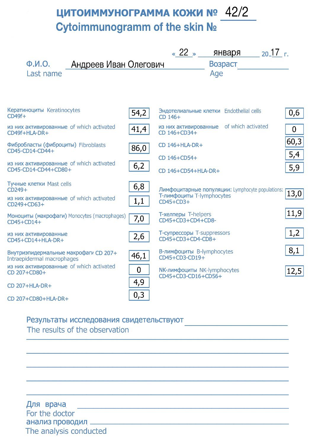
Figure 1. Results of the skin citoimmunogram on a blank sheet
The results of this specific case show that the skin sample contains:
a significant number of keratinocytes, most of which are activated, indicating moderate proliferative activity in the basal layer of the epidermis;
B-lymphocytes, which are normally present in the circulating blood and lymph volume due to their positive taxis towards highly endothelial venules located primarily in lymph nodes. Their presence in the skin indicates the activity of humoral immunity;
several types of T-lymphocytes (CD3+ lymphocytes), which are predominantly located in the three outer layers of the epidermis, and the fact that CD4+ cells outnumber CD8+ cells, indicating an increase in adaptive immunity in the skin;
a low concentration of T-cytotoxic lymphocytes, indicating the absence of an infectious or inflammatory process;
the remaining indicators show the number of specific skin cells, but their activation is low, which, combined with the absence of specific complaints in this patient, indicates a normal condition of his skin.
Observation two is a comparative assessment of the skin citoimmunogram of native and cryopreserved samples
To demonstrate the possibilities of using a skin cytoimmunogram, we selected 64 people and divided them into groups based on gender and age: 25-45 years and 45-65 years, with 16 people in each group. The division of the subjects into gender and age groups was necessary to show the differences in the measured parameters between the different gender and age groups. The study consisted of two stages:
Stage 1: A bioptate from the gluteal area was examined using a patented method.
Stage 2: A cryopreserved analogue of the bioptates was examined.
As a result of the study, for the first time, individual subpopulations were obtained from a heterogeneous population of skin cells, and the phenotype of cells from a native and cryopreserved sample was determined (Table 1).
The viability of the native sample was 99.8%, while the viability of the cryopreserved sample was 87.0%. When comparing the percentage of skin cells in the two samples, no significant differences were found, except for the cells that express HLA-DR antigens and adhesion molecules on their surface, which are known to be antigen-presenting cells.
Table 1. Comparative evaluation of the phenotype of skin biopsy cells from native and cryopreserved samples
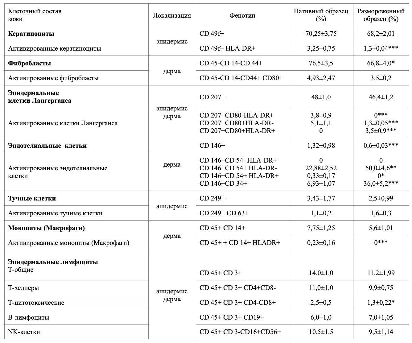
The significance of the difference compared to the native sample * p<0.05; **p<0.01;***p<0.001
Observation three: Comparative analysis of the number of epidermal keratinocytes and dermal fibroblasts
The main components of the skin's immune system are keratinocytes and fibroblasts, which regulate the physiological functions of the skin, and the substances they produce serve as signaling molecules for local coordination of intercellular and intertissue interactions. These subpopulations of skin cells are resident in different layers of the skin, and their coordinated interaction determines their ability to maintain skin homeostasis and respond to external and internal stimuli. As an example, we demonstrate the observation of the number of keratinocytes and fibroblasts in the studied samples (Table 2).
Table 2. Polo-age-related quantitative characteristics of epidermal keratinocytes and dermal fibroblasts of the examined contingent.
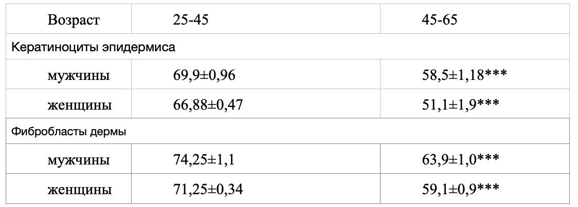
reliability of comparison with the control group: * - p<0,05; ** - p<0,01; *** - p<0,001
As can be seen, the number of keratinocytes in the epidermis decreases over time. The results are reliable in the comparison groups of 25-45 and 45-65 for both sexes, as well as in the comparison groups of 25-45 and 45-65 between men and women. It should be noted that the number of keratinocytes in the epidermis is higher in men than in women by 4%. This can be explained by the fact that the thickness of the skin is significantly greater in men than in women. Moreover, the number of keratinocytes in the epidermis decreases by 16.3% in men and by 23.6% in women with age. This decrease in cells may be due to differences in hormonal levels between the sexes.
The number of fibroblasts in the dermis also demonstrates an age-related decrease. The results are reliable in the comparison groups of 25-45 and 45-65 for both sexes, as well as in the comparison groups of 25-45 and 45-65 between men and women. The number of cells is higher in men than in women by 4%. The dynamics of decrease with age – the number of fibroblasts in the dermis decreases by 13.9% in men and by 17% in women.
There is a greater decrease in the number of keratinocytes in the epidermis than in the number of fibroblasts in the dermis. Moreover, these changes are more pronounced in women.
Discussion
Thus, the proposed method allows us to obtain information about the quantitative composition and functional activity of skin cells, which objectively indicates the current status of the patient's local immunity and can serve as the basis for an individualized treatment and prevention program.
The skin cytoimmunogram, as a method of skin diagnostics, is simple to perform and accessible, allowing us to assess the qualitative and quantitative composition of its individual cell subpopulations, evaluate their function, and assess their response to external and internal environmental factors.
The invention of the skin cytoimmunogram opens up a new understanding of the processes occurring in the human skin for practical dermatology and cosmetology, providing an opportunity to quantify the skin's condition not only within an individual but also within a population, not only in normal conditions but also, importantly, in pathological conditions. Moreover, there is no doubt about the role of the chemokine system in the skin, such as in the pathogenesis of psoriasis [2].
The ability to examine a cryopreserved sample allows for a repeated examination after some time or after certain procedures, treatments, or the use of topical medications or cosmetics, and by comparing the data, the doctor can objectively assess the changes.
The widespread use of the cytoimmunogram in the future will enable the creation of a sex- and age-specific register of skin conditions in both normal and pathological conditions, allowing us to determine the skin's response to environmental factors, measure the activity of native skin cell subpopulations in both normal and pathological conditions, use criteria for age-related skin changes, objectively assess the dynamics of skin diseases, and individually select medications and monitor the effectiveness of topical medications.
«Африка? Как Африка? Тебя не съели? Ну как там? Страшно было? А правда, что у масаев самые длинные... ноги? А ты змею ел? А слона? Слушай, а почему ты там не остался?» — спрашивают меня мои друзья...
After twelve years of successful expeditionary experience, the LIVING PARALLEL broadens the horizon of its search. Since 2019, the program of continuous round-the-WORLD STUDY of the possibilities of Culture as a "second nature" and man in local cultures has been continuing.
Stage 1 has already taken place in New Zealand, and the next stage is ahead – South and Central Africa – 2021

Interview for the author's program "Persona" Anatoly Konstantinovich Omelchuk on the TV channel Russia24 Region-Tyumen.

Оказаться в Лиме для всех участников экспедиции Живая Параллель – знаковое событие, именно здесь стартует исследование закономерностей культуры, о которой, согласитесь, у нас очень смутные представления. А иначе и быть не могло: инки, испанцы, индейцы и... метисы - много разнообразных культур и одна на всех Культура Перу, в которой нам предстоит побыть ближайшие недели.